. 2014:2014:436438.
doi: 10.1155/2014/436438. Epub 2014 Sep 1.

Dendritic cells from aged subjects display enhanced inflammatory responses to Chlamydophila pneumoniae

Affiliations

Dendritic cells from aged subjects display enhanced inflammatory responses to Chlamydophila pneumoniae

Sangeetha Prakash et al. Mediators Inflamm. 2014.

Abstract

Chlamydophila pneumoniae (CPn) is a common respiratory pathogen that causes a chronic and persistent airway infection. The elderly display an increased susceptibility and severity to this infection. However, the underlying mechanisms are not well understood. Dendritic cells (DCs) are the initiators and regulators of immune responses. Therefore, we investigated the role of DCs in the age-associated increased CPn infection in vitro in humans. Though the expression of activation markers was comparable between the two age groups, DCs from aged subjects secreted enhanced levels of proinflammatory mediators such as TNF-α and CXCL-10 in response to CPn. In contrast, the secretion of IL-10 and innate interferons, IFN-α and IFN-λ, was severely impaired in DCs from aged donors. The increased activation of DCs from aged subjects to CPn also resulted in enhanced proliferation of CD4 and CD8 T cells in a DC-T coculture. Furthermore, T cells primed with CPn-stimulated DCs from aged subjects secreted increased levels of IFN-γ and reduced levels of IL-10 compared to DCs obtained from young subjects. In summary, DCs from the elderly displayed enhanced inflammatory response to CPn which may result in airway remodeling and increase the susceptibility of the elderly to respiratory diseases such as asthma.


Figures

Figure 1
Figure 1
Comparable expression of activation markers in DCs from aged and young subjects in response to CPn. (a) Histograms depict the expression of activation markers on DCs from aged and young subjects after on stimulation with CPn. Bar graphs depict the MFI of the expression of activation molecules on CPn stimulated DCs from aged and young subjects. (b) CD80; (c) CD86; (d) CD83; (e) HLADR. Data is mean +/− S.E of 12 aged and 12 young subjects. P value depicted is comparison of CPn-stimulated DCs from aged subjects with their young counterparts.
Figure 2
Figure 2
Altered secretion of cytokines and chemokines by DCs from aged subjects as compared to DCs from young subjects after stimulation with CPn. Graphs depict the levels of cytokine and chemokines secreted by CPn-stimulated DCs from aged and young subjects. (a) CXCL-10; (b) TNF-α; (c) IL-12; (d) IL-10; (e) IFN-α; (f) IFN-λ; (g) IL-6; (h) CXCL-8; (i) IL-1β. Each dot corresponds to a separate subject. Data is of 13 aged and 13 young subjects. P value depicted is comparison of CPn-stimulated DCs from aged subjects with their young counterparts.
Figure 3
Figure 3
Conventional Myeloid DCs (cDCs) from the blood of aged subjects also display enhanced inflammatory responses to CPn. Graphs depict the levels of cytokine and chemokines secreted by CPn-stimulated cDCs purified directly from the blood of aged and young subjects. (a) TNF-α; (b) CXCL-10; (c) IL-6; (d) IL-10; (e) IFN-λ; Each dot corresponds to a separate subject. Data is of 8 aged and 8 young subjects. P value depicted is comparison of CPn-stimulated DCs from aged with their young counterparts.
Figure 4
Figure 4
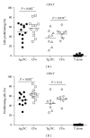
Enhanced proliferation of CD4 and CD8 T cells by CPn-stimulated DCs from aged subjects in an allogeneic DC-T cell coculture. CPn-stimulated and CPn-unstimulated DCs from aged and young subjects were cultured with allogeneic, CFSE labeled T cells from young subjects for 6 days. Graphs depict the proliferation of (a) CD4 T cells; (b) CD8 T cells as determined by flow cytometry. Each dot corresponds to a separate subject. Data is of 12 aged and 9 young subjects for (a) and 11 aged and 8 young for (b).  P value depicted is comparison of CPn-stimulated DCs with unstimulated DCs for both aged and young subjects.
Figure 5
Figure 5
Effect of CPn-stimulated DCs from aged and young donors on T cell cytokine induction. Graphs depict the level of cytokines secreted by T cells after 6 days in an allogeneic coculture with CPn-stimulated DCs from aged and young subjects. (a) IFN-γ; (b) IL-10; (c) IL-17; (d) IL-21. Each dot corresponds to a separate subject. Data is of 10 aged and 10 young subjects. P value depicted is comparison of CPn-stimulated DCs T cell from aged subjects with their young counterparts.

References

    1. Busse PJ, Lurslurchachai L, Sampson HA, Halm EA, Wisnivesky J. Perennial allergen-specific immunoglobulin E levels among inner-city elderly asthmatics. Journal of Asthma. 2010;47(7):781–785. - PubMed
    1. Diaz-Guzman E, Mannino DM. Airway obstructive diseases in older adults: from detection to treatment. Journal of Allergy and Clinical Immunology. 2010;126(4):702–709. - PubMed
    1. Reed CE. Asthma in the elderly: diagnosis and management. Journal of Allergy and Clinical Immunology. 2010;126(4):681–687. - PubMed
    1. Matsuse H, Tsuchida T, Fukahori S, et al. Differential airway inflammatory responses in asthma exacerbations induced by respiratory syncytial virus and influenza virus A. International Archives of Allergy and Immunology. 2013;161(4):378–382. - PubMed
    1. McKenna JJ, Bramley AM, Skarbinski J, Fry AM, Finelli L, Jain S. Asthma in patients hospitalized with pandemic influenza A(H1N1)pdm09 virus infection-United States, 2009. BMC Infectious Diseases. 2013;13(1, article 57) - PMC - PubMed