. 2014 Sep 20;14(1):95.
doi: 10.1186/s12935-014-0095-7. eCollection 2014.

MicroRNA-492 expression promotes the progression of hepatic cancer by targeting PTEN

Affiliations

MicroRNA-492 expression promotes the progression of hepatic cancer by targeting PTEN

Jianxin Jiang et al. Cancer Cell Int. .

Abstract

Background: Aberrant microRNA (miRNA) expression plays an essential role in the pathogenesis of Hepatocellular Carcinoma (HCC). However, specific involvement of miRNAs in HCC remains incompletely understood. The aim of this study was to explore the relevant microRNAs involved in the development of HCC.

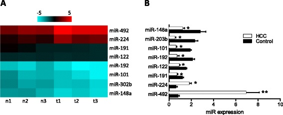
Methods: MicroRNA microarray was used to screen for the differentially expressed miRNAs in cancerous tissue and adjacent non-cancerous control tissue from patients with HCC (n = 3). Quantitative PCR was subsequently used to verify the results of microarray. Based on the findings, we investigated the role of miR-492 in the pathogenesis of HCC in vitro and in vivo using three tumor cells lines. Furthermore, we analyzed the clinical correlation of miR-492 expression with patient survival (n = 28).

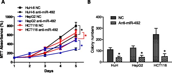
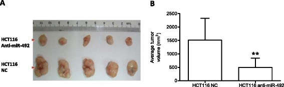
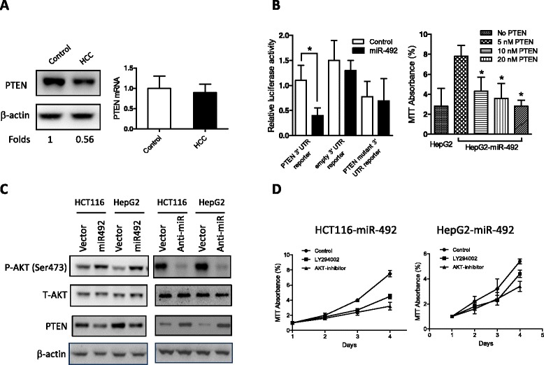
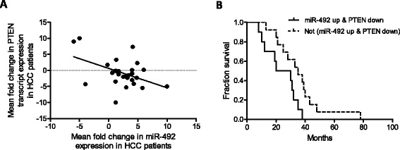
Results: We showed that microRNA-492 (miR-492) was elevated in HCC samples from patients with hepatic cancer. Knockdown of miR-492 attenuated the proliferation of cancer cell lines in vitro and inhibited primary tumor growth in vivo in SCID mice. We identified PTEN as a functional target for miR-492. Overexpression of miR-492 resulted in decreased PTEN expression and was associated with increased AKT activation in cancer cell lines. Moreover, miR-492-mediated increase of the proliferation of cancer cells was able to be suppressed by a PI3K inhibitor and an AKT inhibitor. The HCC patients with high miR-492/low PTEN had poorer survival.

Conclusions: miR-492 is implicated in the regulation of HCC progression through PTEN and AKT pathway. The data suggest that miR-492 is a biomarker of HCC and a potential therapeutic target for hepatocellular carcinoma.

Keywords: AKT; Hepatocellular carcinoma; PTEN; microRNA.


Figures

Figure 1
Figure 1
Comparison of miRNA expression profile between HCC tissues and adjacent non-cancerous tissues using an array-based miRNA profiling. (A) The heat map diagram shows the result of the differentially expressed miRNAs (only differentially expressed miRNAs with fold change > =1.5 or fold change = <0.8 and p value <0.05 are included) from miRNA microarray. (B) The expression levels of these miRNAs were measured by qRT-PCR and normalized to the expression of U6 in each sample. Data are the mean ± SD (n = 3) of one representative experiment. Similar results were obtained in three independent experiments. n: adjacent non-cancerous tissues, t: HCC tissues. *refers to p < 0.05 and **refers to p < 0.01 in a Student’s t test between the HCC group and the control group.
Figure 2
Figure 2
Knockdown of miR-492 attenuates cencer cell proliferation and colony formation in vitro. (A) The expression of miR-492 was inhibited using siRNA transfection to cancer cell lines HuH-6, HCT116 and HepG2. Subsequently, the proliferations of cancer cells were determined using MTT. Statistical analysis was carried out on day 4 and day 5. (B) Colony formation of three cell lines were measured in the culture treated with siRNA to miR-492 and control siRNA (NC). Data are presented as mean ± SD. *refers to p < 0.05 between the groups.
Figure 3
Figure 3
Anti-miR-492 inhibits the in vivo growth of HCT116 in SCID mice . (A) HCT116 cells (5 x 10⁶) were infected with lentiviral siRNAs to miR-492 or scramble control and subsequently transferred into SCID mice (n = 5 per group). After 30 days, tumors were isolated from mice and their volume were calculated as “volume = length × width2 × π/6”. (B) The average tumor volume was calculated from 5 mice per group and statistically analyzed to compare the difference between the experimental group and the control group. **refers to p < 0.01.
Figure 4
Figure 4
PTEN is a functional target of miR-492 in HCCs . (A) HCC tissues and the adjacent non-cancerous tissues were subject to Western blot and real-time PCR analyses to assess PTEN mRNA and protein levels. Data are the mean ± SD (n = 4) of one representative experiment. Similar results were obtained in at least three independent experiments. (B) HEK-293 cells were transfected with either miR-492 mimics (20 nM) or the mutant 3′-UTR of miR-492 (20 nM) along with the matched control using 80 ng of indicated vectors and 40 ng of pRL-TK. After 24 h, firefly luciferase activity was measured and normalized by Renilla luciferase activity. Data are the mean ± SD (n = 4) of one representative experiment. Similar results were obtained in at least three independent experiments. The proliferation of HepG2 cells transfected with miR-492 plasmid (0.5 nM) and different concentrations of PTEN plasmid (5 nM, 10 nM, 20 nM) was determined by MTT. *indicates the significant difference between the control and PTEN transfected cells. (C) HCT116 or HepG2 cells were infected with the indicated lentivirus for 24 h, and p-AKT, total AKT and PTEN levels were detected by immunoblot analysis. Data shown is one representative experiment out of three. (D) miR-492 transfection enhanced the proliferation of both HCT116 and HepG2 which was inhibited by the PI3K and ATK inhibitors.
Figure 5
Figure 5
Upregulation of miR-492 and downregulation of PTEN expression is associated with poorer survival of HCC patients . (A) Scatter plot represents the statistically significant correlation in the relative transcript expression of miR-492 and PTEN (p < 0.001) in the tumor versus paired adjacent non-tumor tissues from 28 HCC patients. Each spot represents data from one HCC patient presented in the Log2 scale and the linear regression line is marked as the solid line. (B) Kaplan-Meier survival curve for HCC patients classified based on whether HCC tumor showed miR-492 upregulation (more than 2 fold) and PTEN downregulation (more than 1.5 fold), compared to the paired adjacent non-tumor samples. Upregulation of miR-492 and downregulation of PTEN expression is significantly associated with poorer patient survival (p <0.05).

References

    1. Bushati N, Cohen SM. microRNA functions. Annu Rev Cell Dev Biol. 2007;23:175–205. doi: 10.1146/annurev.cellbio.23.090506.123406. - DOI - PubMed
    1. Sicard F, Gayral M, Lulka H, Buscail L, Cordelier P. Targeting miR-21 for the therapy of pancreatic cancer. Mol Ther J Am Soc Gene Therapy. 2013;21(5):986–994. doi: 10.1038/mt.2013.35. - DOI - PMC - PubMed
    1. Park SM, Gaur AB, Lengyel E, Peter ME. The miR-200 family determines the epithelial phenotype of cancer cells by targeting the E-cadherin repressors ZEB1 and ZEB2. Genes Dev. 2008;22(7):894–907. doi: 10.1101/gad.1640608. - DOI - PMC - PubMed
    1. Lowery AJ, Miller N, Dwyer RM, Kerin MJ. Dysregulated miR-183 inhibits migration in breast cancer cells. BMC Cancer. 2010;10:502. doi: 10.1186/1471-2407-10-502. - DOI - PMC - PubMed
    1. Wang B, Hsu SH, Wang X, Kutay H, Bid HK, Yu J, Ganju RK, Jacob ST, Yuneva M, Ghoshal K. Reciprocal regulation of microRNA-122 and c-Myc in hepatocellular cancer: role of E2F1 and transcription factor dimerization partner 2. Hepatology. 2014;59(2):555–566. doi: 10.1002/hep.26712. - DOI - PMC - PubMed

LinkOut - more resources