. 2014 Sep 25;10(9):e1004406.
doi: 10.1371/journal.ppat.1004406. eCollection 2014 Sep.

Persistence of virus reservoirs in ART-treated SHIV-infected rhesus macaques after autologous hematopoietic stem cell transplant

Affiliations

Persistence of virus reservoirs in ART-treated SHIV-infected rhesus macaques after autologous hematopoietic stem cell transplant

Maud Mavigner et al. PLoS Pathog. .

Abstract

Despite many advances in AIDS research, a cure for HIV infection remains elusive. Here, we performed autologous hematopoietic stem cell transplantation (HSCT) in three Simian/Human Immunodeficiency Virus (SHIV)-infected, antiretroviral therapy (ART)-treated rhesus macaques (RMs) using HSCs collected prior to infection and compared them to three SHIV-infected, ART-treated, untransplanted control animals to assess the effect of conditioning and autologous HSCT on viral persistence. As expected, ART drastically reduced virus replication, below 100 SHIV-RNA copies per ml of plasma in all animals. After several weeks on ART, experimental RMs received myeloablative total body irradiation (1080 cGy), which resulted in the depletion of 94-99% of circulating CD4+ T-cells, and low to undetectable SHIV-DNA levels in peripheral blood mononuclear cells. Following HSC infusion and successful engraftment, ART was interrupted (40-75 days post-transplant). Despite the observed dramatic reduction of the peripheral blood viral reservoir, rapid rebound of plasma viremia was observed in two out of three transplanted RMs. In the third transplanted animal, plasma SHIV-RNA and SHIV DNA in bulk PBMCs remained undetectable at week two post-ART interruption. No further time-points could be assessed as this animal was euthanized for clinical reasons; however, SHIV-DNA could be detected in this animal at necropsy in sorted circulating CD4+ T-cells, spleen and lymph nodes but not in the gastro-intestinal tract or tonsils. Furthermore, SIV DNA levels post-ART interruption were equivalent in several tissues in transplanted and control animals. While persistence of virus reservoir was observed despite myeloablation and HSCT in the setting of short term ART, this experiment demonstrates that autologous HSCT can be successfully performed in SIV-infected ART-treated RMs offering a new experimental in vivo platform to test innovative interventions aimed at curing HIV infection in humans.


Conflict of interest statement

The authors have declared that no competing interests exist.

Figures

Figure 1
Figure 1. Experimental design.
(A) Three RMs received G-CSF 50 mg/kg subcutaneously daily for six consecutive days prior to HSC collection by leukopheresis and cryopreservation of the collected cells. Two apheresis procedures were performed on each transplant recipient. After collection of pre-infection HSCs, RMs were infected i.v. with 10,000 TCID50 RT-SHIVTC. Starting at day 28 post-infection, RMs received ART daily. After 37 to 54 days on ART, the three experimental RMs underwent TBI (total dose of 1080 cGy), fractionated in three doses given on three consecutive days pre-transplant. On the two following days, the leukopheresis products were infused. ART was interrupted 40 to 75 days post-transplant. (B) Three control RMs were infected i.v. with 10,000 TCID50 RT-SHIVTC and received ART for the same period of time as in the transplanted animals. The three control RMs did not undergo TBI/autologous HSCT.
Figure 2
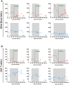
Figure 2. Virologic and immunologic characteristics pre-transplant.
(A) The levels of SHIV-RNA, expressed as copies/ml of plasma are shown for each individual animal. Dotted line represents the limit of detection of the assay. (B) Longitudinal assessment of the absolute numbers of circulating CD4+ T-cell expressed as cells per µl. Transplanted animals are depicted in red and controls in blue. Shaded area represents the period of ART treatment.
Figure 3
Figure 3. Autologous hematopoietic stem cell transplant.
(A) Comparison of the pre- and post-transplant absolute numbers of circulating neutrophils, monocytes, lymphocytes and CD4+ T-cell expressed as cells per µl. Pre-transplant time point is the final assessment prior to TBI (SHIV-infected, on ART). Post-transplant time point represents the nadir cell count observed during the eleven days following TBI. Longitudinal assessment of the absolute numbers of circulating (B) neutrophils and (C) platelets expressed as cells per µl. Transplanted animals are depicted in red, controls in blue. Shaded area represents the period of ART treatment. Yellow area represents the period of platelet transfusion support. The dotted lines indicate the minimum level of neutrophils or platelets used to define engraftment.
Figure 4
Figure 4. Effect of autologous HSCT on white blood cell and CD4+ T-cell counts.
Longitudinal assessment of the absolute numbers of white blood cells (A) and circulating CD4+ T-cells (B) expressed as cells per µl are shown for each individual animal. Transplanted animals are depicted in red, controls in blue. Shaded area represents the period of ART treatment.
Figure 5
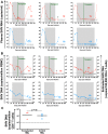
Figure 5. SHIV-RNA and -DNA levels post ART interruption.
(A) Longitudinal assessment of the level of SHIV-RNA, expressed as copies/ml of plasma. (B) Longitudinal assessment of cell associated SHIV-DNA. Plain lines represent the level of SHIV-DNA in PBMCs determined by PCR. The dashed lines represent the estimated level of SHIV-DNA in CD4+ T-cells calculated based on PBMC frequency of infection determined by PCR and the frequency of CD4 + T-cells in PBMC determined by flow cytometry. Shaded area represents the period of ART treatment. (C) SHIV-DNA levels determined by PCR at necropsy, in sorted peripheral CD4+ T-cells and expressed as copies/million cells. Lines are drawn at the geometric mean. Mann Whitney U test was used to determine significance. Transplanted animals are depicted in red, controls in blue. Grey dotted lines represent the limit of detection of the assay.
Figure 6
Figure 6. SHIV-DNA in tissues post ART interruption.
SHIV-DNA levels expressed as copies/million cells obtained at necropsy from the ileum, jejunum, colon, rectum, superficial and mesenteric lymph nodes, and tonsils are shown for each individual animal. Transplanted animals are depicted in red, controls in blue. Estimated number of CD4+ T-cells per million cells in tissues at necropsy is indicated in Table S1.

Similar articles

Cited by

References

    1. Chun TW, Stuyver L, Mizell SB, Ehler LA, Mican JA, et al. (1997) Presence of an inducible HIV-1 latent reservoir during highly active antiretroviral therapy. Proc Natl Acad Sci U S A 94: 13193–13197. - PMC - PubMed
    1. Finzi D, Hermankova M, Pierson T, Carruth LM, Buck C, et al. (1997) Identification of a reservoir for HIV-1 in patients on highly active antiretroviral therapy. Science 278: 1295–1300. - PubMed
    1. Chun TW, Justement JS, Murray D, Hallahan CW, Maenza J, et al. (2010) Rebound of plasma viremia following cessation of antiretroviral therapy despite profoundly low levels of HIV reservoir: implications for eradication. AIDS 24: 2803–2808. - PMC - PubMed
    1. Davey RT Jr, Bhat N, Yoder C, Chun TW, Metcalf JA, et al. (1999) HIV-1 and T cell dynamics after interruption of highly active antiretroviral therapy (HAART) in patients with a history of sustained viral suppression. Proc Natl Acad Sci U S A 96: 15109–15114. - PMC - PubMed
    1. Hutter G, Nowak D, Mossner M, Ganepola S, Mussig A, et al. (2009) Long-term control of HIV by CCR5 Delta32/Delta32 stem-cell transplantation. N Engl J Med 360: 692–698. - PubMed

Publication types

MeSH terms