. 2014 Sep 25;10(9):e1004368.
doi: 10.1371/journal.ppat.1004368. eCollection 2014 Sep.

Hematopoietic but not endothelial cell MyD88 contributes to host defense during gram-negative pneumonia derived sepsis

Affiliations

Hematopoietic but not endothelial cell MyD88 contributes to host defense during gram-negative pneumonia derived sepsis

Miriam H P van Lieshout et al. PLoS Pathog. .

Abstract

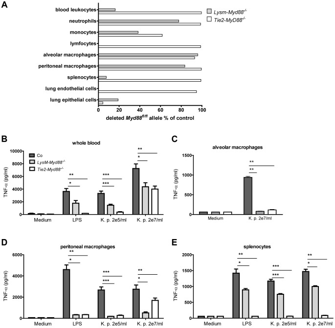
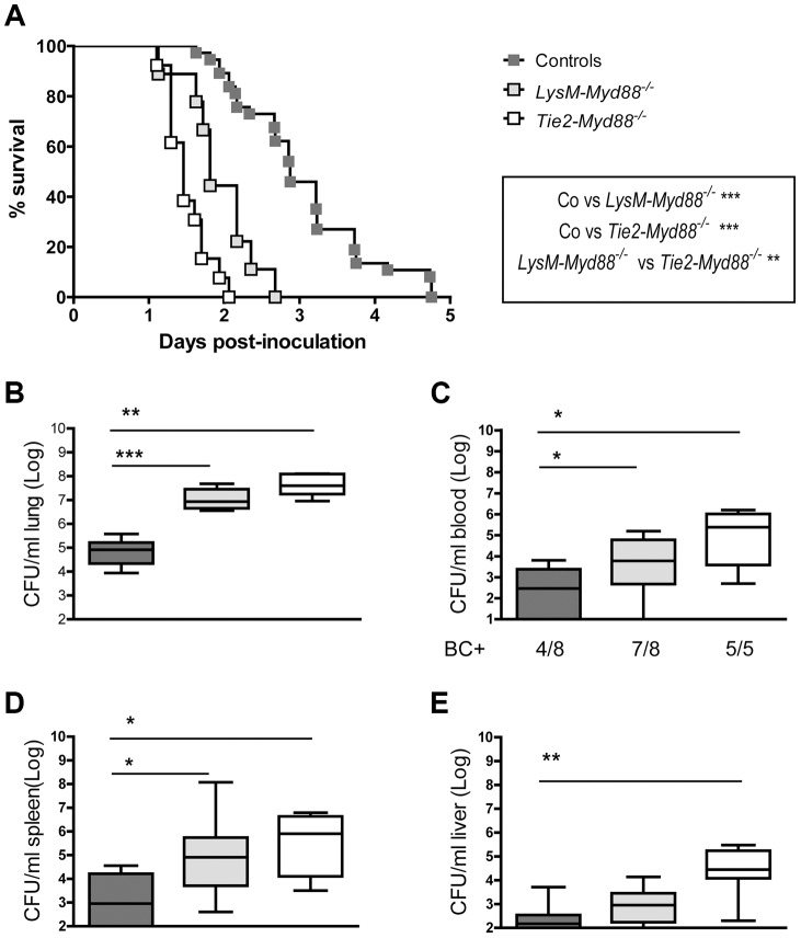
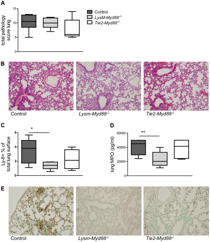
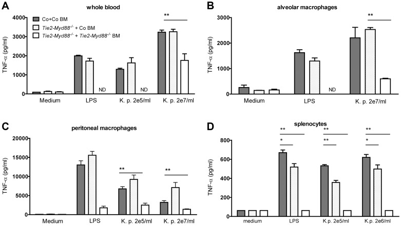
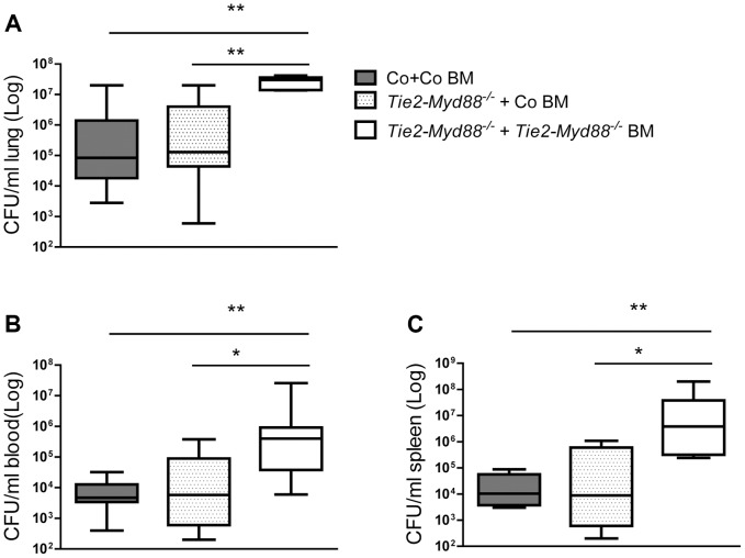
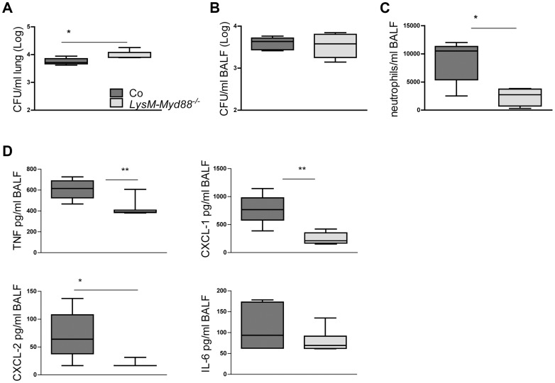
Klebsiella pneumoniae is an important cause of sepsis. The common Toll-like receptor adapter myeloid differentiation primary response gene (MyD)88 is crucial for host defense against Klebsiella. Here we investigated the role of MyD88 in myeloid and endothelial cells during Klebsiella pneumosepsis. Mice deficient for MyD88 in myeloid (LysM-Myd88(-/-)) and myeloid plus endothelial (Tie2-Myd88(-/-)) cells showed enhanced lethality and bacterial growth. Tie2-Myd88(-/-) mice reconstituted with control bone marrow, representing mice with a selective MyD88 deficiency in endothelial cells, showed an unremarkable antibacterial defense. Myeloid or endothelial cell MyD88 deficiency did not impact on lung pathology or distant organ injury during late stage sepsis, while LysM-Myd88(-/-) mice demonstrated a strongly attenuated inflammatory response in the airways early after infection. These data suggest that myeloid but not endothelial MyD88 is important for host defense during gram-negative pneumonia derived sepsis.


Conflict of interest statement

The authors have declared that no competing interests exist.

Figures

Figure 1
Figure 1. Genetic and functional characterization of primary cells from LysM-Myd88−/− and Tie2-Myd88−/− mice.
The residual amount of the MyD88fl/fl allel in blood and primary cells LysM-Myd88−/− and Tek-Myd88−/− mice was quantified via qRT-PCR relative to the unaffected Socs-3 gene. The amount of remaining “floxed” MyD88 region in LysM-MyD88−/− and Tek-MyD88−/− mice was calculated using the 2-deltaCt (ΔΔCt) method using the amount of genomic DNA from Myd88fl/fl mice for the no-deletion control. The deletion efficiency was calculated as (1 - residual Myd88fl) ×100% (A). Whole blood (B), alveolar and peritoneal macrophages (C,D) and splenocytes (E) derived from control, LysM-Myd88−/− and Tie2-Myd88−/− mice (n = 3 per group) were in vitro stimulated with LPS derived from Klebsiella pneumoniae (1 µg/ml) or heat killed K. pneumoniae in two concentrations (2×10⁵ CFU/ml or 2×10⁷/ml) for 20 hours. Data are expressed as mean (SE). * p<0.05, ** p<0.01, *** p<0.001.
Figure 2
Figure 2. Impaired survival and bacterial defense in LysM-Myd88−/− and Tie2-Myd88−/− mice.
Control, LysM-Myd88−/− and Tie2-Myd88−/− mice were intranasally infected with ∼6×10³ CFU K. pneumoniae. Survival of control (dark grey symbols, n = 37), LysM-Myd88−/− (light grey symbols, n = 9) and Tie2-Myd88−/− mice (white symbols, n = 13) expressed as Kaplan-Meier plot (A), bacterial loads in lung (B), blood (C), spleen (D) and liver (E), of control (dark grey bars, n = 8), LysM-Myd88−/− (light grey bars, n = 8) and Tie2-Myd88−/− mice (white bars, n = 5 mice). Data are expressed as box-and-whisker diagrams depicting the smallest observation, lower quartile, median, upper quartile, and largest observation. BC+ = number of positive blood cultures. Survival curves were compared with Log-Rank test Bacterial loads were compared to control mice determined with Mann-Whitney U test: * p<0.05, ** p<0.01, *** p<0.001.
Figure 3
Figure 3. Lung inflammatory response.
Mice were intranasally infected with ∼6×10³ CFU K. pneumoniae; Histological scores 24 hours after infection determined as described in the Methods section, in control (dark grey, n = 8), LysM-Myd88−/− (light grey, n = 8) and Tie2-Myd88−/− mice (white, n = 5) (A). Panel (B) shows representative lung histology of control, LysM-Myd88−/− and Tie2-Myd88−/− mice H&E staining, original magnification 20×. Neutrophil influx compared between mouse groups as reflected by Ly6 lung surface positivity (C) and whole lung MPO levels (D). Panel (E) shows representative images of Ly-6 staining on lung slides from control, LysM-Myd88−/− and Tie2-Myd88−/− mice; Data are expressed as box-and-whisker diagrams depicting the smallest observation, lower quartile, median, upper quartile, and largest observation. * p<0.05, ** p<0.01.
Figure 4
Figure 4. Bone marrow transfer restores responsiveness of hematopoietic cells from Tie2-Myd88−/− mice to Klebsiella.
Whole blood (A), alveolar and peritoneal macrophages (B,C) and splenocytes (D) derived from control mice transplanted with control bone marrow (Co+ Co BM, grey bars) and Tie2-Myd88−/− mice transplanted with control bone marrow (Tie2-Myd88−/−+Co BM, white dotted bars) or Tie2-Myd88−/− bone marrow (Tie2-Myd88−/−+Tie2-Myd88−/− BM, white bars). (n = 2–3 per group) were in vitro stimulated with LPS derived from Klebsiella pneumoniae (1 µg/ml) or heat killed K. pneumoniae in two concentrations (2×10⁵ CFU/ml or 2×10⁷/ml) for 20 hours. Data are expressed as mean (SE). * p<0.05, ** p<0.01. ND = not determined.
Figure 5
Figure 5. The absence of MyD88 in the hematopoietic compartment determines the impaired antibacterial defense of Tie2-Myd88−/− mice.
Control and Tie2-Myd88−/− mice were irradiated and injected with control or Tie2-Myd88−/− bone marrow cells. Six weeks after transplantation, mice were infected with 6×10³ CFU K. pneumoniae and sacrificed after 24 hours. Bacterial loads in lung (A), blood (B), spleen (C) of control mice transplanted with control bone marrow (Co+ Co BM, grey bars, n = 8) and Tie2-Myd88−/− mice transplanted with control bone marrow (Tie2-Myd88−/−+Co BM, white dotted bars) or Tie2-Myd88−/− bone marrow (Tie2-Myd88−/−+Tie2-Myd88−/− BM, white bars). Data are expressed as box-and-whisker diagrams depicting the smallest observation, lower quartile, median, upper quartile, and largest observation. * p<0.05, ** p<0.01.
Figure 6
Figure 6. LysM-Myd88−/− mice demonstrate an attenuated early inflammatory response.
Control and LysM-Myd88−/− mice were intranasally infected with ∼6×10³ CFU K. pneumoniae. Bacterial loads in lung (A) and BALF (B), number of neutrophils (C) and levels of TNF-α, CXCL-1, CXCL-2 and IL-6 (D) in BALF of control (dark grey symbols, n = 8) and LysM-Myd88−/− mice (light grey symbols, n = 8). Data are expressed as box-and-whisker diagrams depicting the smallest observation, lower quartile, median, upper quartile, and largest observation. * p<0.05, ** p<0.01.

Similar articles

Cited by

References

    1. World Health Organisation (2012) WHO causes of death 2008, Global health Observatory, World Health Organisation.
    1. Kollef MH, Shorr A, Tabak YP, Gupta V, Liu LZ, et al. (2005) Epidemiology and outcomes of health-care-associated pneumonia: results from a large US database of culture-positive pneumonia. Chest 128: 3854–3862. - PubMed
    1. Welte T, Torres A, Nathwani D (2012) Clinical and economic burden of community-acquired pneumonia among adults in Europe. Thorax 67: 71–79 thx.2009.129502 [pii];10.1136/thx.2009.129502 [doi] - DOI - PubMed
    1. Zahar JR, Timsit JF, Garrouste-Org, Francais A, Vesin A, et al. (2011) Outcomes in severe sepsis and patients with septic shock: pathogen species and infection sites are not associated with mortality. Crit Care Med 39: 1886–1895 10.1097/CCM.0b013e31821b827c [doi] - DOI - PubMed
    1. Coque TM, Baquero F, Canton R (2008) Increasing prevalence of ESBL-producing Enterobacteriaceae in Europe. Euro Surveill 13 19044. - PubMed

Publication types

MeSH terms