. 2014 Sep 25;9(9):e108680.
doi: 10.1371/journal.pone.0108680. eCollection 2014.

Oligomerisation of C. elegans olfactory receptors, ODR-10 and STR-112, in yeast

Affiliations

Oligomerisation of C. elegans olfactory receptors, ODR-10 and STR-112, in yeast

Muhammad Tehseen et al. PLoS One. .

Abstract

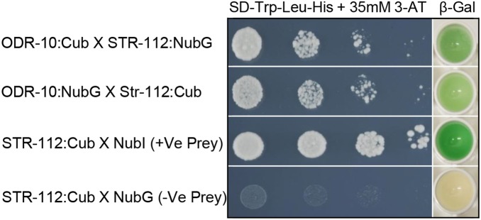
It is widely accepted that vertebrate G-Protein Coupled Receptors (GPCRs) associate with each other as homo- or hetero-dimers or higher-order oligomers. The C. elegans genome encodes hundreds of olfactory GPCRs, which may be expressed in fewer than a dozen chemosensory neurons, suggesting an opportunity for oligomerisation. Here we show, using three independent lines of evidence: co-immunoprecipitation, bioluminescence resonance energy transfer and a yeast two-hybrid assay that nematode olfactory receptors (ORs) oligomerise when heterologously expressed in yeast. Specifically, the nematode receptor ODR-10 is able to homo-oligomerise and can also form heteromers with the related nematode receptor STR-112. ODR-10 also oligomerised with the rat I7 OR but did not oligomerise with the human somatostatin receptor 5, a neuropeptide receptor. In this study, the question of functional relevance was not addressed and remains to be investigated.


Conflict of interest statement

Competing Interests: The authors have declared that no competing interests exist.

Figures

Figure 1
Figure 1. Yeast cells expressing ODR-10-GFP2.
Arrows indicate the plasma membrane and cytoplasmic localisation. Bar: 11.9 µm. Image was obtained with a Leica SP2 confocal laser scanning microscope using excitation at 488 nm.
Figure 2
Figure 2. Three lines of evidence for formation of ODR-10 homo-oligomers.
A. Co-immunoprecipitation of a pair of tagged chemoreceptor subunits. B. ODR-10-Rluc co-expressed with SSTR5-GFP2 as a negative control. Following membrane extraction with digitonin and immunoprecipitation with an anti-GFP2 antibody, as described in Materials and Methods, luciferase activity was measured using 5 µM Coelenterazine H with an emission filter of 475±30 nm. Values represent means ± SD of experiments performed on three independent transformants (**denotes significance at P≤0.001), “b” is significantly different from “a”. C. BRET2 ratios measured to test for homo-dimerisation of ODR-10 in intact yeast cells. Values represent means ± SD (n = 3) (**denotes significance at P≤0.001), “b” is significantly different from “a”. Indicated constructs in both A and C were expressed separately, or mixed (+), or co-expressed (/). D. ODR-10 homo-oligomerises in the split-ubiquitin yeast two-hybrid system. The C-terminal half of ubiquitin (Cub) was fused to the C-terminus of the full-length cDNA of odr-10 (ODR-10::Cub). The N-terminal half of ubiquitin (NubG) was fused to the C- termini of full-length cDNA of odr-10 (ODR-10::NubG). The interaction of ODR-10::Cub with pOST1:NubI served as a positive control to ensure the correct topology of the fusion protein. The interaction of ODR-10::Cub with the empty vector (pPR3-STE) served as a negative control. Yeast transformants containing both a Cub fusion and a NubG fusion construct were grown on drop out media (SD -Leu and -Trp) (Figure S2A) and selective medium lacking histidine (SD -Leu, -Trp and -His) containing 35 mM 3-Amino-1, 2, 4-triazole (3-AT). β-galactosidase assays were performed to verify interactions. Cells were spotted as one-tenth dilutions starting at Abs600nm = 1. E. ODR-10 does not hetero-oligomerise with SSTR5 in the split-ubiquitin yeast two-hybrid system. Growth and β-galactosidase activity of yeast cells expressing ODR-10:Cub and SSTR5:NubG fusions. The interaction of SSTR5::NubG with the empty vector (pBT3-STE) served as a negative control. Yeast transformants containing both a Cub fusion and a NubG fusion constructs were grown on drop out media (SD -Leu and -Trp) (Figure S2E) and selective medium lacking histidine (SD -Leu, -Trp and -His) containing 35 mM 3-Amino-1, 2, 4-triazole (3-AT). β-Galactosidase assays were performed to verify interactions. Cells were spotted as one-tenth dilutions starting at Abs600nm = 1.
Figure 3
Figure 3. Constructs used for split-ubiquitin assays.
A. Bait fusion proteins were made with the C-terminus of ubiquitin (Cub) linked to the synthetic transcription factor LexA-VP16 at the C-termini of ODR-10, STR-112 and I7 in the pBT3-STE plasmid. The STE2 sequence improves translation of protein constructs. B. Prey protein fusions were constructed with the mutant N-terminal part of ubiquitin (NubG) at the C termini of the SSTR5, STR-112, ODR-10 and I7. If bait and prey interact, NubG and Cub are brought into close proximity, resulting in the reconstitution of functional ubiquitin and release of the LexA-VP16 transcriptional factor, which leads to transcriptional readout, resulting in growth of host yeast cells on the selective medium (SD-L-T-H) or color development in a β-galactosidase assay.
Figure 4
Figure 4. ODR-10 hetero-oligomerises with STR-112 in the split-ubiquitin yeast two-hybrid system.
Growth and β-Galactosidase activity of yeast cells expressing various combinations of Cub and NubG fusions. The control plasmids were pPR3-STE and pBT3-STE. Yeast transformants containing both Cub fusion and NubG fusion constructs were grown on drop out media (SD -Leu and -Trp) (Figure S2B) and selective medium lacking histidine (SD -Leu, -Trp and -His) containing 35 mM 3-Amino-1, 2, 4-triazole (3-AT). β-galactosidase assays were performed to verify interactions. Cells were spotted as one-tenth dilutions starting at Abs600nm = 1.
Figure 5
Figure 5. ODR-10 hetero-oligomerises with I7.
A. Co-immunoprecipitation of pairs of tagged chemoreceptor subunits. Values represent means ± SD of experiments performed in triplicate with three independent transformants for each condition (**denotes significance at P≤0.001), “b” is significantly different from “a”. B. BRET2 ratios measured to test for hetero-dimerisation of ODR-10 and I7 in intact yeast cells. Values represent means ± SD (n = 3) (**denotes significance at P≤0.002), “b” is significantly different from “a”. Indicated constructs in both A and B were expressed separately, or mixed (+), or co-expressed (/). C. ODR-10 hetero-oligomerises with I7 in the split-ubiquitin yeast two-hybrid system. Growth and β-galactosidase activity of yeast cells expressing ODR-10:Cub and I7:NubG fusions. Yeast transformants containing both Cub fusion and NubG fusion constructs were grown on drop out media (SD -Leu and -Trp) (Figure S2C) and selective medium lacking histidine (SD -Leu, -Trp and -His) containing 35 mM 3-Amino-1, 2, 4-triazole (3-AT). β-galactosidase assays were performed to verify interactions. Cells were spotted as one-tenth dilutions starting at Abs600nm = 1.
Figure 6
Figure 6. I7 forms homo-oligomers.
A. Co-immunoprecipitation of a pair of tagged chemoreceptor subunits. Values represent means ± SD of experiments performed in triplicate with three independent transformants for each condition (**denotes significance at P≤0.001), “b” is significantly different from “a”. B. BRET2 ratios measured to test for homo-oligomerisation of I7 in intact yeast cells. Values represent means ± SD of experiments performed in triplicate with three independent transformants for each condition. (*denotes significance at P≤0.01). Indicated constructs in both A and B were expressed separately, or mixed (+), or co-expressed (/). C. I7 homo-oligomerises in the split-ubiquitin yeast two-hybrid system. Growth and β-galactosidase activity of yeast cells expressing I7:Cub and I7:NubG fusions. The interaction of I7::Cub with the empty vector (pPR3-STE) served as a negative control. Yeast transformants containing both a Cub fusion and a NubG fusion constructs were grown on drop out media (SD -Leu and -Trp) (Figure S2D) and selective medium lacking histidine (SD -Leu, -Trp and -His) containing 35 mM 3-Amino-1,2,4-triazole (3-AT). β-galactosidase assays were performed to verify interactions. Cells were spotted as one-tenth dilutions starting at Abs600nm = 1.

References

    1. Gurevich VV, Gurevich EV (2008) How and why do GPCRs dimerize? Trends in Pharmacological Sciences 29: 234–240. - PMC - PubMed
    1. Marinissen MJ, Gutkind JS (2001) G-protein coupled receptors and signaling networks: emerging paradigms. Trends in Pharmacological Sciences 22: 368–376. - PubMed
    1. Jordan BA, Trapaidze N, Gomes I, Nivarthi R, Devi LA (2001) Oligomerisation of opioid receptors with beta 2-adrenergic receptors: a role in trafficking and mitogen-activated protein kinase activation. Proc Natl Acad Sci USA 98: 343–348. - PMC - PubMed
    1. George SR, O’Dowd BF, Lee SP (2002) G-protein coupled receptor oligomerisation and its potential for drug discovery. Nat Rev Drug Discov 1: 808–820. - PubMed
    1. Kroeger KM, Pfleger KD, Eidne KA (2003) G-protein coupled receptor oligomerisation in neuroendocrine pathways. Front Neuroendocrinol 24: 254–278. - PubMed

Publication types

MeSH terms

Substances

LinkOut - more resources