. 2014 Sep 25;10(9):e1004374.
doi: 10.1371/journal.ppat.1004374. eCollection 2014 Sep.

Reorganization of the endosomal system in Salmonella-infected cells: the ultrastructure of Salmonella-induced tubular compartments

Affiliations

Reorganization of the endosomal system in Salmonella-infected cells: the ultrastructure of Salmonella-induced tubular compartments

Viktoria Krieger et al. PLoS Pathog. .

Abstract

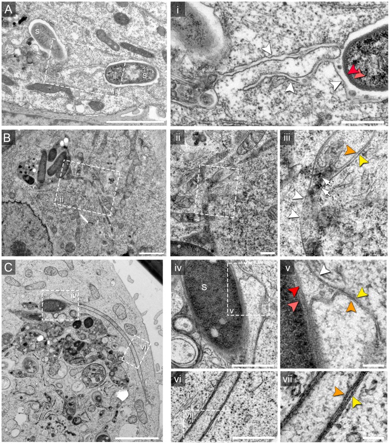
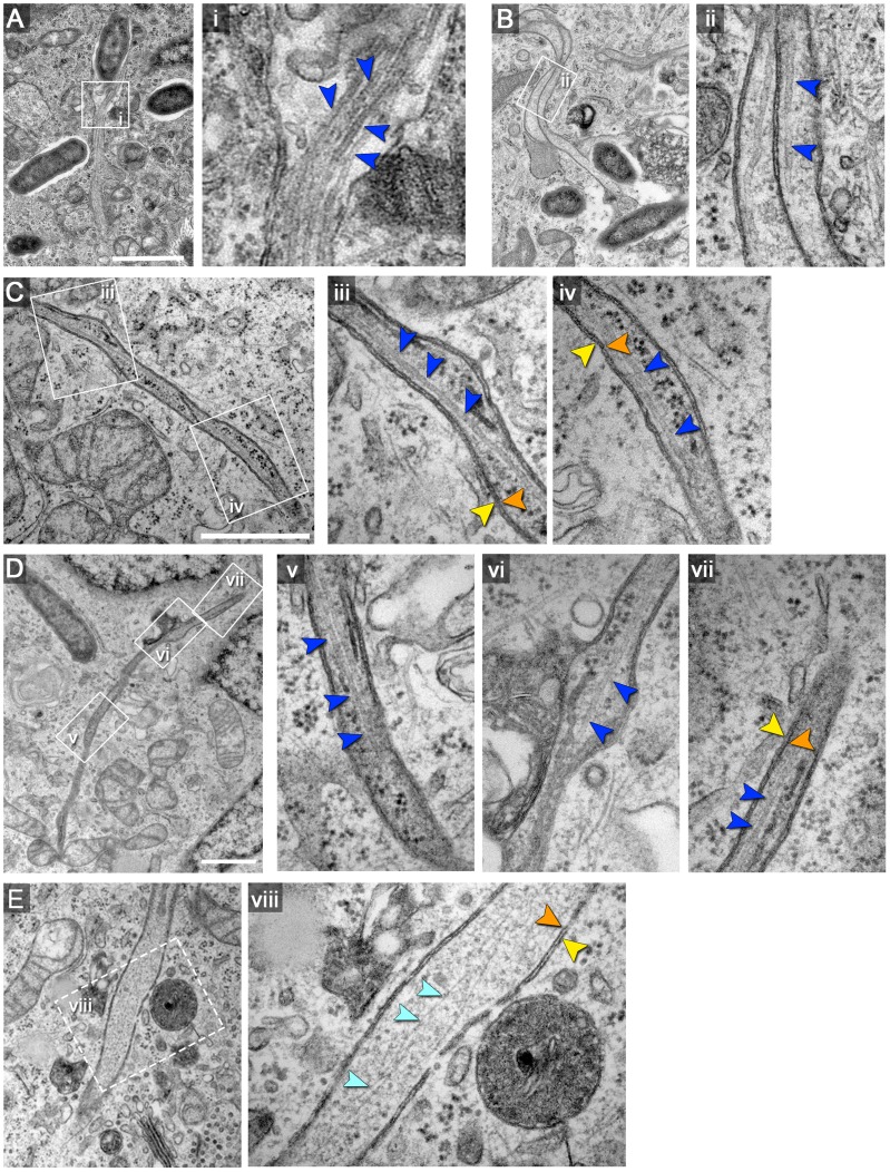
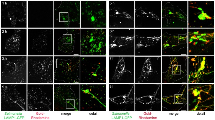
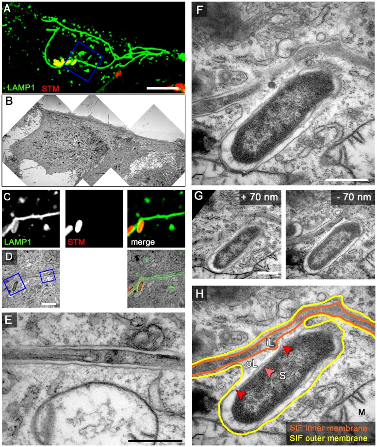
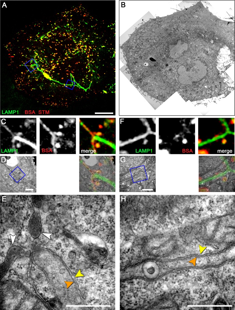
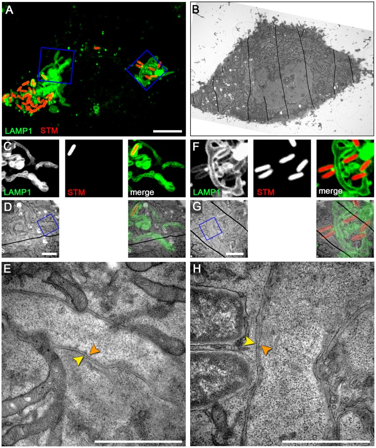
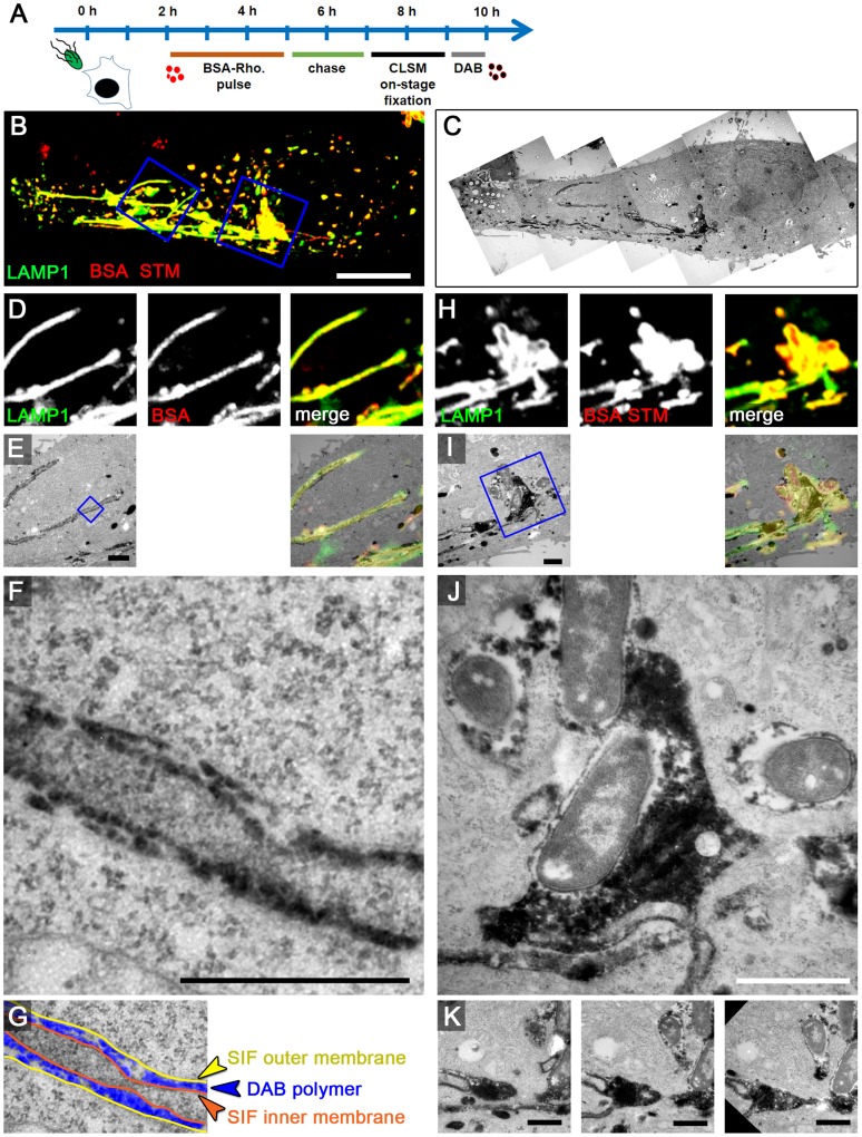
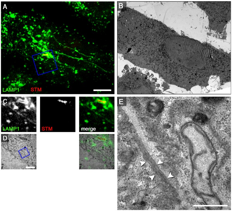
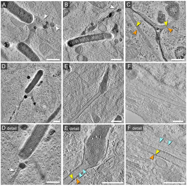
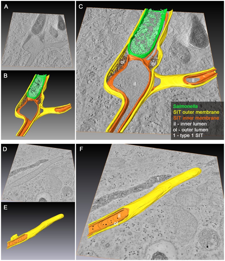
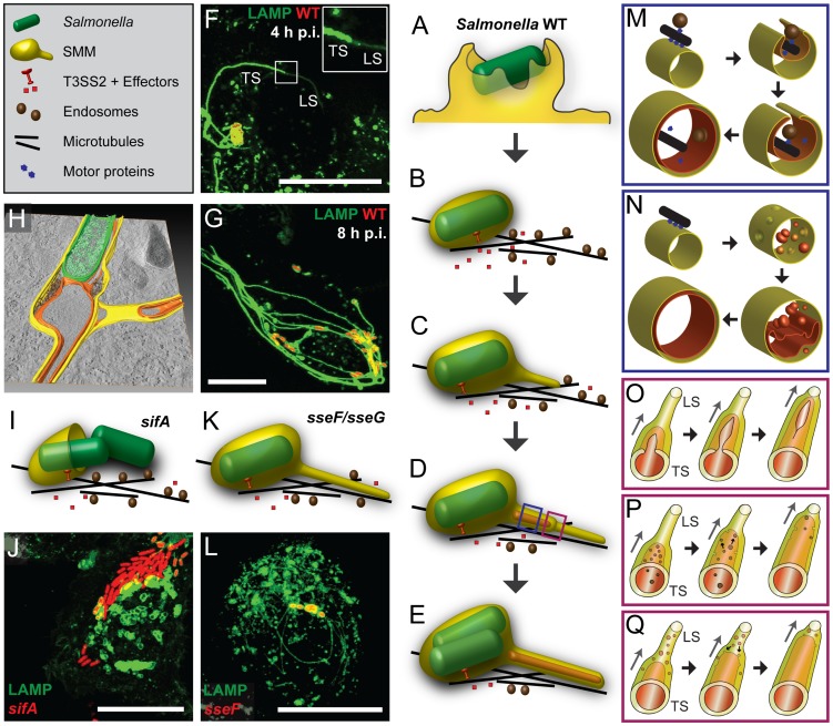
During the intracellular life of Salmonella enterica, a unique membrane-bound compartment termed Salmonella-containing vacuole, or SCV, is formed. By means of translocated effector proteins, intracellular Salmonella also induce the formation of extensive, highly dynamic membrane tubules termed Salmonella-induced filaments or SIF. Here we report the first detailed ultrastructural analyses of the SCV and SIF by electron microscopy (EM), EM tomography and live cell correlative light and electron microscopy (CLEM). We found that a subset of SIF is composed of double membranes that enclose portions of host cell cytosol and cytoskeletal filaments within its inner lumen. Despite some morphological similarities, we found that the formation of SIF double membranes is independent from autophagy and requires the function of the effector proteins SseF and SseG. The lumen of SIF network is accessible to various types of endocytosed material and our CLEM analysis of double membrane SIF demonstrated that fluid phase markers accumulate only between the inner and outer membrane of these structures, a space continual with endosomal lumen. Our work reveals how manipulation of the endosomal membrane system by an intracellular pathogen results in a unique tubular membrane compartmentalization of the host cell, generating a shielded niche permissive for intracellular proliferation of Salmonella.


Conflict of interest statement

The authors have declared that no competing interests exist.

Figures

Figure 1
Figure 1. Single and double membrane SIT extend from the SCV in Salmonella-infected cells.
Electron micrographs of ultrathin sections through HeLa cells infected with Salmonella enterica wild type (S). Cells were subjected to HPF-FS at 10 h post infection (p.i.). Representative micrographs of SCV and SIT are shown. A) Example of thin single-membrane SIT interconnecting two SCV B) Cells were pre-loaded with BSA-coupled 10 nm colloidal gold particles to track lumen of endosomes and luminal space of SCV/SIT. Micrograph shows an example of SCV connected to a single-membrane SIT. The SIT lumen is filled with electron-dense content and BSA-gold particles (black dots indicated by white arrows) (iii). Note that parts of SIT appear as double membrane compartments. C) Double membrane tubular compartment emerging from the SCV. Details of membrane organization of SCV (iv, v) and SIT (vi, vii). Note that central portions of SIT have density identical to the cytosol. White arrowheads indicate single membrane compartments and for double membrane compartments, orange and yellow arrowheads distinguish inner and outer membrane, respectively. Light and dark red arrowheads indicate inner and outer membrane of the Salmonella cell envelope, respectively. Scale bars: 2 µm (A, B, C), 500 nm (i, ii, iv, vi), 100 nm (iii, v, vii).
Figure 2
Figure 2. Double membrane SIT contain cytoskeletal filaments and ribosomes.
HeLa cells were infected with S. enterica WT as for Figure 1. A), B), C) and D) show examples of double-membrane SIT with microtubules, indicated by dark blue arrowheads. Orange and yellow arrowheads indicate inner and outer membranes of SIT, respectively. Measurements of microtubules as shown in i) revealed an average diameter of 22 nm±2.3 nm. Higher magnifications are shown in panel ii to vii. E) shows an example of a double-membrane SIT containing thinner, actin-like filaments (light blue arrowheads). Note that the inner space of all double-membrane SIT (A–E) also contains ribosomes. Scale bars: 500 nm.
Figure 3
Figure 3. The SIF network is accessible to various types of endosomal cargo.
HeLa cells expressing LAMP1-GFP (green) were infected with Salmonella expressing GFP (green, rod-shaped structures). Pulse-chase experiments were performed by addition of 10 nm gold Nanoparticles conjugated with BSA-Rhodamine (red) at various time points prior and after infection. Live cell imaging of infected cells was performed and representative cells are shown. LAMP1-GFP-positive tubules emerged at 3 to 4 h p.i. Note the almost complete co-localization of endosomal cargo with tubular, LAMP1-GFP-positive SIT. Scale bar: 10 µm.
Figure 4
Figure 4. LAMP1-GFP-positive SIF display a double membrane at 8 h p.i.
HeLa cells expressing LAMP1-GFP (green) were seeded in Petri dishes with a gridded coverslip and infected with Salmonella expressing mCherry (STM, red). Live cell imaging was performed 8 h p.i. to visualize LAMP1-GFP-positive SIF (A, maximum intensity projection [MIP], C, single Z plane). Subsequently, the cells were fixed and processed for CLEM to reveal the ultrastructure of selected cells. Several low magnification images were stitched to visualize the cell morphology (B). Higher magnification images were used to align LM and TEM images (D). Details of a LAMP1-GFP-positive double membrane SIF (E) and an SCV linked to a SIF (F, G) are shown. G) Two additional ultrathin sections show the membrane organization of the SCV and SIF in detail F). H) The inner and outer membrane of a SIF and SCV are outlined in orange and yellow, respectively. Light and dark red arrowheads indicate inner and outer membrane of the Salmonella cell envelope, respectively. Labels: S, Salmonella; M, mitochondria; iL, inner lumen; oL, outer lumen. A cell representative for 10 biological replicates is shown (1–3 technical replicates with each 2–4 cells). Scale bars: 10 µm (A, B), 2 µm (C, D), 500 nm (E, F, G).
Figure 5
Figure 5. Early-stage SIF in HeLa cells exhibit leading and trailing phenotypes.
HeLa expressing LAMP1-GFP (green) were seeded on a Petri dish with gridded coverslip and infected with Salmonella WT expressing mCherry (STM, red). After infection, cells were pulse-chased with BSA-Rhodamine (red) 1–3 h p.i. Live cell imaging was performed 4 h p.i. to visualize LAMP1-GFP-positive SIF (A, MIP; C, F, single Z plane). Subsequently, the cells were fixed and processed for CLEM. Several low magnification images were stitched to visualize the cell morphology (B). C–H) CLEM of ROIs showing SIF of various diameter (D, E), and a double membrane SIF with an internal vesicle (G, H). E) A tubular compartment showing the transition from a leading SIF with a single membrane and endosomal content to a trailing SIF with a double membrane and cytosolic content. H) Double membrane SIF with luminal membrane vesicle. Orange and yellow arrowheads indicate inner and outer membranes if double membrane SIF, respectively. The white arrowheads indicate single membrane SIF. A cell representative for three biological replicates is shown (1–3 technical replicates with each 2–4 cells). Scale bars: 10 µm (A, B), 1 µm (C, D, E, G), 500 nm (E, H).
Figure 6
Figure 6. RAW264.7 macrophages infected with Salmonella exhibit double membrane SIF.
RAW264.7 cells expressing LAMP1-GFP (green) were seeded in Petri dishes with a gridded coverslip and infected with Salmonella WT expressing mCherry (STM, red). Live cell imaging was performed 12 h p.i. to visualize LAMP1-GFP-positive SIF (A, MIP; C, F, single Z plane). Subsequently, the cells were fixed and processed for CLEM to reveal the ultrastructure. Several low magnification images were stitched to visualize the cell morphology (B). Higher magnification images were used to align LM and TEM images (D, G). Details of LAMP1-GFP-positive double membrane SIF (E, H) are shown. A cell representative of three biological replicates is shown (1–2 technical replicates with each 2–4 cells). Scale bars: 10 µm (A, B), 2 µm (C, D), 500 nm (E).
Figure 7
Figure 7. The outer lumen of double membrane SIF is in interchange with endocytosed material.
A) Scheme of the experiment. HeLa expressing LAMP1-GFP (green) were seeded on a petri dish with a gridded coverslip. Cells were infected with Salmonella WT expressing mCherry (STM, red). BSA-Rhodamine was added as fluid tracer to the medium 2–5 h p.i. After live cell imaging of selected cells at 8 h p.i. by CLSM (B, MIP), cells were immediately fixed on stage. DAB photo-conversion by Rhodamine was performed and cells were prepared for TEM. Several images of the same section were stitched for an overview (C). Details for LAMP1-positive, fluid tracer-labeled SIF (D–G) and SCV with attached SIF (H–K) are shown by correlative live cell CLSM (D, H, single Z plane) and TEM (E–G, I–K) micrographs. F) shows a higher magnification of double membrane SIF, and the pseudocolored micrograph (G) indicates the organization of inner and outer SIF membrane and the DAB polymer deposition in intermembrane lumen. J) shows DAP polymer deposition in direct contact to Salmonella within the SCV. Successive sections with several SIF extending from the SCV are shown in K). A cell representative of three biological replicates is shown (1–3 technical replicates with each 2–4 cells). Scale bars: 10 µm (B, C), 1 µm (D, E, H, I, K), 500 nm (F, J).
Figure 8
Figure 8. The SPI2-T3SS effector SseF is required for induction of double membrane SIF.
For CLEM analysis, HeLa cells stably transfected with LAMP1-GFP (green) were seeded on a Petri dish with a gridded coverslip and infected with the Salmonella sseF-deficient strain expressing mCherry (STM, red). After live cell imaging at 8 h p.i. by CLSM (A, MIP) cells were fixed immediately on stage. sseF-infected HeLa cells exhibit thin LAMP1-positive tubules. B) Low magnification TEM micrograph of the same cell. Details for LAMP1-positive thin SIF and Salmonella within SCV are shown by correlative live cell CLSM (C, F single Z plane) and TEM (D, G) micrograph. E, H) Higher magnifications of a SIF. The single membrane tubule is indicated by arrowheads. A cell representative of two biological replicates is shown (1–2 technical replicates with each 2–4 cells). Scale bars: 10 µm (A, B), 2 µm (C, D, F, G), 500 nm (E, H).
Figure 9
Figure 9. Morphology of SCV and SIT analyzed by EM tomography.
HeLa cells were infected with Salmonella WT as before, fixed and processed for EM tomography of partial cell volumes. A) to F) shows representative images corresponding to the tilt series shown in Movies S2, S3, S4, S5, S6, S7, S8, S9, S10. Note the presence of double membrane SIT indicated by orange and yellow arrowheads. SIT that are delimited by single membranes and that contain multi-lamellar vesicles and dense granules typical for late endosomes and lysosomes are referred to as type 1 SIT (A, B, D), with white arrowheads indicating SIT membranes. SIT that appear delimited by double membranes and lack multi-vesicular membranes are referred to as type 2 SIT (E, F). C) shows a ‘hybrid’ SIT resulting from partial fusion of two intertwined type 1 and type 2 SIT. E) shows a type 2 SIT with an inner space filled by a bundle of actin-like filaments, while SIT in F) are associated with F-actin filaments adjacent to the SIT (light blue arrowheads). Scale bars: 500 nm.
Figure 10
Figure 10. 3D organization of SCV and SIT in Salmonella-infected cells.
HeLa cells were infected with Salmonella WT for 10 h and processed for EM tomography of partial cell volumes followed by 3D-surface rendering. Representative images of single and double tilt series are shown in A) and D). A–C) Example of an SCV with extending and branching type 2 SIT showing dense luminal content between the two adjacent membranes at the base of SIT. D–F) Example of type 2 SIT with a closed tip and two membranes that wrap up portion of ribosome-containing cytosol. Note the presence of an adjacent type 1 SIT (without 3D-surface rendering) with a dense luminal content and delimited by a single membrane (E). The corresponding tilt series are shown in Movie S11 and Movie S13, respectively. B) and E) show 3D-surface rendering of the SIT and SCV membrane organization representing Movie S12 and Movie S14. Inner and outer SIT membranes are indicated in orange and yellow, respectively. The resulting inner and outer lumen of the double membrane tubular structure are labeled with iL and oL, respectively. Merged TEM images and rendered models are shown in C) and F).
Figure 11
Figure 11. Models for the biogenesis of SIF.
Schematic depiction of events leading to engulfment of cytosol and cytoskeletal elements, and formation of double membrane structures. After invasion of host cells by Salmonella involving ruffle formation (A), the internalized pathogen is located in a host-derived membrane compartment, called Salmonella-containing vacuole (SCV) that moves along microtubules (B) towards the MTOC. Subsequently, Salmonella activates the SPI2-T3SS and translocation of effector proteins into the host cytosol, thereby starting the induction of SIF. These tubular structures are formed through fusion processes with endosomes that move along microtubules (MT). In the initial phase (3–4 h p.i.) SIF consist only of one membrane (C) that undergo a process resulting in SIT consisting of double membranes (D, E). In the initial phase of centrifugal extension of SIT (D, F), we observed thinner single membrane SIF at the tips (leading SIF, LS) and thicker double membrane SIF at the base (trailing SIF, TS). At later time points (8–10 h p.i.) a stable double membrane SIF network is induced by Salmonella WT, accompanied by a strong reduction of host cell endosomes (E, G). The tomogram of a Salmonella WT-infected cell indicates the features of SCV and double membrane SIT (H). The sifA mutant strain is not capable of maintaining the membrane integrity of the SCV, Salmonella escape from the damaged SCV into the cytoplasm and no SIF formation is observed (I, J). The sseF and sseG mutant strains induce thin SIF consisting of one membrane (K, L). M) and N) show scenarios for conversion of single to double membrane SIF (blue boxes). M) Tubular membranes are formed along MT. By lateral extension, these membranes enwrap the guiding MT, as well as vesicles transported along MT and portions of cytoplasm. Membrane fusions results in formation of double membrane SIF. N) Alternatively, membrane invagination generates small vesicles that accumulate within single membrane SIF and the fusion of these vesicles results in formation of inner membrane tubule and its continuous extension. In both models, the lumen of the inner tubule is segregated from Salmonella. O), P), Q) Models for the dynamic conversion of LS to TS (red boxes). See main text for further details. Scale bars: 20 µm.

References

    1. Schaible UE, Haas A, editors (2009) Intracellular Niches for Microbes: A Pathogens Guide Through the Host Cell. Weinheim: Wiley-VCH.
    1. Figueira R, Holden DW (2012) Functions of the Salmonella pathogenicity island 2 (SPI-2) type III secretion system effectors. Microbiology 158: 1147–1161. - PubMed
    1. Ibarra JA, Steele-Mortimer O (2009) Salmonella - the ultimate insider. Salmonella virulence factors that modulate intracellular survival. Cell Microbiol 11: 1579–1586. - PMC - PubMed
    1. Garcia-del Portillo F, Finlay BB (1995) Targeting of Salmonella typhimurium to vesicles containing lysosomal membrane glycoproteins bypasses compartments with mannose 6-phosphate receptors. J Cell Biol 129: 81–97. - PMC - PubMed
    1. Salcedo SP, Holden DW (2003) SseG, a virulence protein that targets Salmonella to the Golgi network. EMBO J 22: 5003–5014. - PMC - PubMed

Publication types

MeSH terms

Substances