. 2014 Oct;9(10):2476-92.
doi: 10.1038/nprot.2014.167. Epub 2014 Sep 25.

Generation of cell-based systems to visualize chromosome damage and translocations in living cells

Affiliations

Generation of cell-based systems to visualize chromosome damage and translocations in living cells

Vassilis Roukos et al. Nat Protoc. 2014 Oct.

Abstract

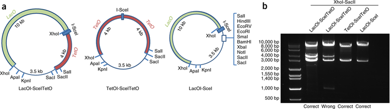
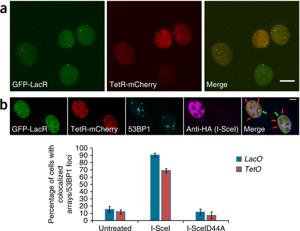
Traditional methods for the generation of DNA damage are not well suited for the observation of spatiotemporal aspects of damaged chromosomal loci. We describe a protocol for the derivation of a cellular system to induce and to visualize chromosome damage at specific sites of the mammalian genome in living cells. The system is based on the stable integration of endonuclease I-SceI recognition sites flanked by bacterial LacO/TetO operator arrays, coupled with retroviral-mediated integration of their fluorescent repressors (LacR/TetR) to visualize the LacO/TetO sites. Expression of the I-SceI endonuclease induces double-strand breaks (DSBs) specifically at the sites of integration, and it permits the dynamics of damaged chromatin to be followed by time-lapse microscopy. Sequential LacO-I-SceI/TetO-I-SceI integrations in multiple chromosomes permit the generation of a system to visualize the formation of chromosome translocations in living cells. This protocol requires intermediate cell culture and molecular biology skills, and it is adaptable to the efficient derivation of any integrated clonal reporter system of interest in ≈ 3-5 months.


Conflict of interest statement

COMPETING FINANCIAL INTERESTS The authors declare no competing financial interests.

Figures

Figure 1 |
Figure 1 |
Overview of the protocol. The cell line of interest is sequentially transfected with the Tet0I-ScelTet0 and Lac0I-Scel vectors together with plasmids conferring resistance to antibiotics, and cell clones containing both integrations are isolated (Steps 18–36). Stable cell lines that emerge are transduced with retroviral vectors expressing fluorescent versions of the LacR (green) and TetR (red) repressors (Steps 37–51), and clones are selected on the basis of optimal LacR/TetR expression detected by microscopy (Steps 52–56), from top to bottom: cells with overabundant LacR expression but optimal TetR expression (green nucleus, red dot), cells with optimal LacR and TetR expression (light yellow nucleus, green and red dots), cells with overabundant LacR and TetR expression (bright yellow nucleus, no dots visible) and cells with overabundant TetR expression but optimal LacR expression (red nucleus, green dot). The selected clones are tested for their ability to induce DSBs by colocalization analysis of the Lac0/Tet0 arrays with the recruitment of a repair protein (blue dot) after the expression of the endonuclease I-SceI (Steps 57–76) and used to assess DSB dynamics (Steps 77–86).
Figure 2 |
Figure 2 |
Characterization of the Lac0/Tet0 plasmids. (a) Maps of the plasmids containing arrays of the Lac0 operators (256× repeats, 10 kb) and the Tet0 operators (96× repeats, 4 kb) adjacent to the restriction site of the endonuclease I-SceI used to generate cell lines to visualize DSBs. (b) Restriction fragment analysis using the XhoI and SacII restriction enzymes, used to verify the integrity of the arrays in the plasmids.
Figure 3 |
Figure 3 |
Selection of clones containing various numbers of array integrations. (a) Isolated clones with one, two or three Tet0I-SceITet0 integrations (arrows). The Tet0 operators were visualized by transient expression of the TetR repressor tagged with mCherry. Nuclei were stained with Hoechst. Scale bars, 10 μm. (b) A clone with three Tet0I-SceITet0 (red) and a single Lac0I-SceI (green) integration. Lac0 and Tet0 operators were visualized by transient expression of the GFP-LacR and TetR-mCherry repressors. Nuclei were stained with Hoechst. Scale bar, 10 μm.
Figure 4 |
Figure 4 |
A cell-based system to visualize chromosome damage in living cells. (a) The Lac0/Tet0 operator/repressor system marks distinct genome sites of integrated recognition sequences of the endonuclease I-SceI. Clones with stable expression of the GFP-LacR and TetR-mCherry repressors, optimal for detection by microscopy, are isolated. (b) Cells left untreated or transfected with the endonuclease I-SceI or the inactive form I-SceID44A for 12 h were fixed and immunofluorescence was performed to detect cells that were positive for I-SceI/I-SceID44A expression (anti-HA staining, magenta) and to visualize 53BP1 repair foci (cyan). The percentage of cells with colocalization of the Lac0 arrays (green arrows) and Tet0 arrays (red arrows) with 53BP1 repair foci was calculated and plotted. Scale bars, 10 μm.
Figure 5 |
Figure 5 |
High-throughput time-lapse microscopy to follow DSB dynamics. Cells described in Figure 4a were transfected with the I-SceI plasmid, and 6 h later time-lapse microscopy was performed to follow DSBs marked by the Lac0 (green dots) and Tet0 (red dots) for up to 20 h. Maximal projected image sequences are shown. Scale bar, 10 μm.

Similar articles

Cited by

References

    1. Ciccia A & Elledge SJ The DNA damage response: making it safe to play with knives. Mol Cell. 40, 179–204 (2010). - PMC - PubMed
    1. Misteli T & Soutoglou E The emerging role of nuclear architecture in DNA repair and genome maintenance. Nat. Rev. Moi. Ceil Biol. 10, 243–254 (2009). - PMC - PubMed
    1. Soria G, Polo SE & Almouzni G Prime, repair, restore: the active role of chromatin in the DNA damage response. Mol. Cell 46, 722–734 (2012). - PubMed
    1. Dion V & Gasser SM Chromatin movement in the maintenance of genome stability. Cell 152, 1355–1364 (2013). - PubMed
    1. Jakob B, Splinter J, Durante M & Taucher-Scholz G Live cell microscopy analysis of radiation-induced DNA double-strand break motion. Proc. Natl. Acad. Sci. USA 106, 3172–3177 (2009). - PMC - PubMed

Publication types

MeSH terms

Substances