. 2014 Sep 25;5(9):e1426.
doi: 10.1038/cddis.2014.386.

MiR-630 inhibits proliferation by targeting CDC7 kinase, but maintains the apoptotic balance by targeting multiple modulators in human lung cancer A549 cells

Affiliations

MiR-630 inhibits proliferation by targeting CDC7 kinase, but maintains the apoptotic balance by targeting multiple modulators in human lung cancer A549 cells

J-X Cao et al. Cell Death Dis. .

Abstract

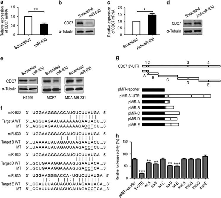
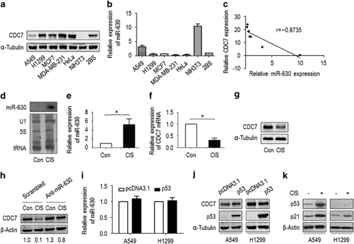
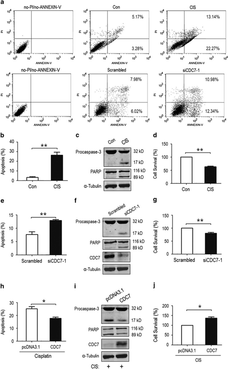
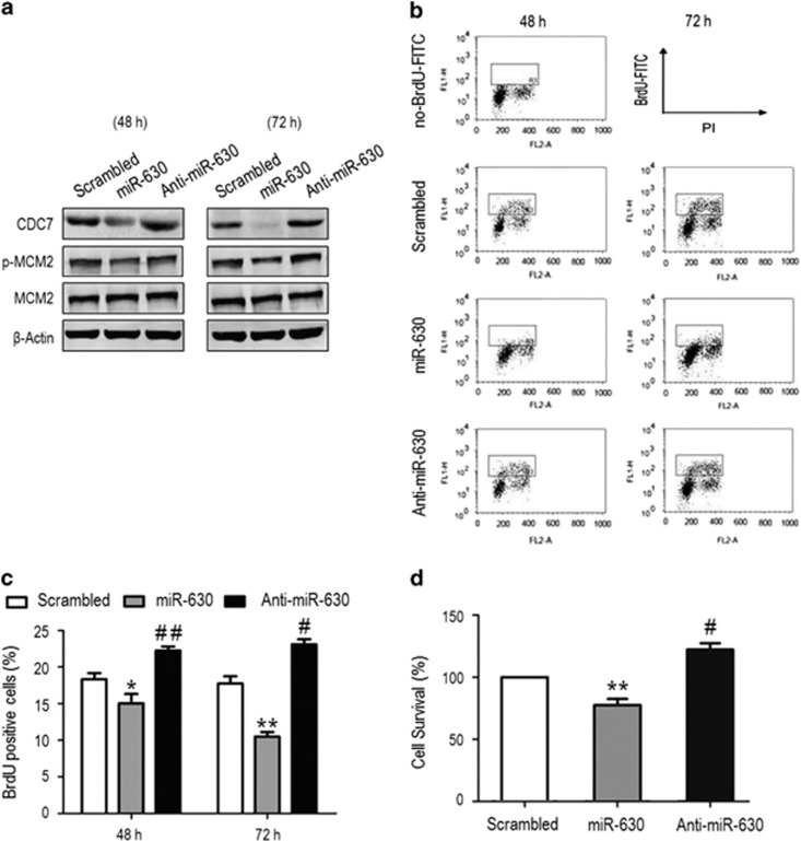
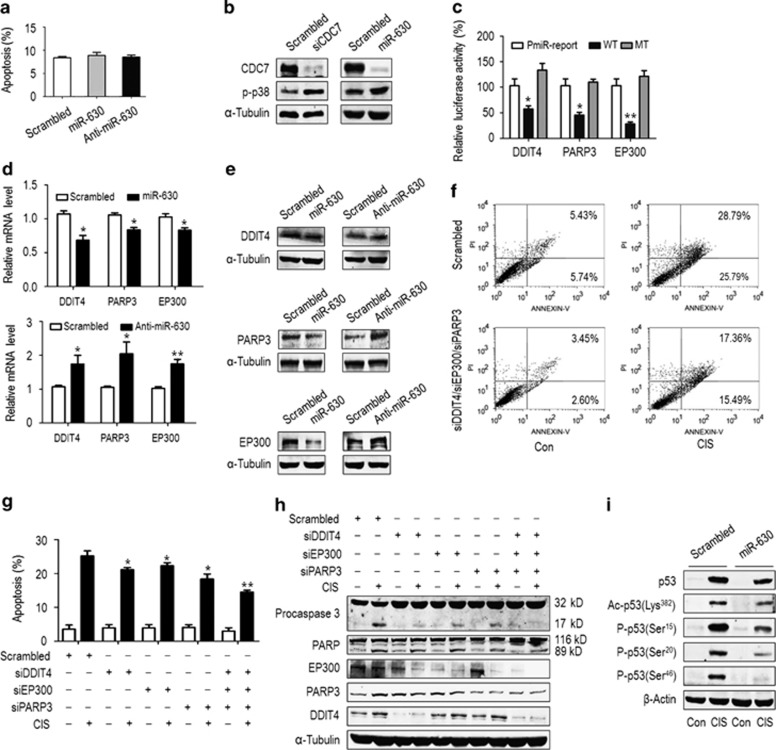
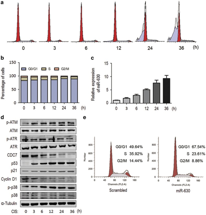
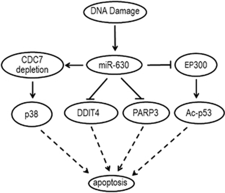
MicroRNAome analyses have shown microRNA-630 (miR-630) to be involved in the regulation of apoptosis. However, its apoptotic role is still debated and its participation in DNA replication is unknown. Here, we demonstrate that miR-630 inhibits cell proliferation by targeting cell-cycle kinase 7 (CDC7) kinase, but maintains the apoptotic balance by targeting multiple activators of apoptosis under genotoxic stress. We identified a novel regulatory mechanism of CDC7 gene expression, in which miR-630 downregulated CDC7 expression by recognizing and binding to four binding sites in CDC7 3'-UTR. We found that miR-630 was highly expressed in A549 and NIH3T3 cells where CDC7 was downregulated, but lower in H1299, MCF7, MDA-MB-231, HeLa and 2BS cells where CDC7 was upregulated. Furthermore, the induction of miR-630 occurred commonly in a variety of human cancer and immortalized cells in response to genotoxic agents. Importantly, downregulation of CDC7 by miR-630 was associated with cisplatin (CIS)-induced inhibitory proliferation in A549 cells. Mechanistically, miR-630 exerted its inhibitory proliferation by blocking CDC7-mediated initiation of DNA synthesis and by inducing G1 arrest, but maintains apoptotic balance under CIS exposure. On the one hand, miR-630 promoted apoptosis by downregulation of CDC7; on the other hand, it reduced apoptosis by downregulating several apoptotic modulators such as PARP3, DDIT4, EP300 and EP300 downstream effector p53, thereby maintaining the apoptotic balance. Our data indicate that miR-630 has a bimodal role in the regulation of apoptosis in response to DNA damage. Our data also support the notion that a certain mRNA can be targeted by several miRNAs, and in particular an miRNA may target a set of mRNAs. These data afford a comprehensive view of microRNA-dependent control of gene expression in the regulation of apoptosis under genotoxic stress.


Figures

Figure 1
Figure 1
MiR-630 downregulates CDC7 expression by targeting CDC7 3'-UTR. A549 cells were transfected with miR-630 mimic or an inhibitor (50 nM) for 48 h. (a) RT-qPCR for CDC7 mRNA downregulation by miR-630. CDC7 mRNA was quantified by the 2−ΔΔCt method, in which glyceraldehyde 3-phosphate dehydrogenase (GAPDH) was used as an internal control. The scrambled small interfering RNA (siRNA) was used as a negative control, under which condition the level of CDC7 mRNA was normalized to ‘1'. Data are presented as mean±S.D. (n=3). (b) Western blotting for CDC7 protein downregulation by miR-630. α-Tubulin was used as a loading control. (c) RT-qPCR and (d) western blotting for effects of miR-630 inhibitor on CDC7 mRNA and protein expression. Anti-scrambled siRNA was used as a control. Data are presented as mean±S.D. (n=3). (e) Western blotting for CDC7 protein in H1299, MCF7 and MDA-MB-231 cells transfected with miR-630 mimic for 48 h. (f) The predicted miR-630-binding sequences or mutated versions of CDC7 3'-UTR fragments ‘A', ‘B', ‘D' and ‘E' in (g). WT, wild type; MT, mutant (mutated bases are underlined). (g) Interpretation of luciferase reporter plasmids containing full-length CDC7 3'-UTR, fragments ‘A' to ‘E' or mutants (upper panel). The full-length 3'-UTR, truncates ‘A' to ‘E' and mutants in (f) were inserted into the pMIR-Report plasmid to generate pMIR-Report-PmiR-3'-UTR and its variations. ‘1' to ‘4' indicates the miR-630 binding sites. (h) Relative luciferase activities of the reporter plasmids in A549 cells. Data are presented as mean±S.D. (n=3). *P<0.05, **P<0.01 and ***P<0.001
Figure 2
Figure 2
MiR-630 expression inversely correlates with CDC7 expression in a variety of cells. (a) Western blotting for CDC7 protein in A549, H1299, MCF7, MDA-MB-231, HeLa, NIH3T3 and 2BS cells. Cells were grown in culture for 48 h. Western blotting was performed with anti-CDC7 antibody. α-Tubulin was used as a loading control. (b) Stem-loop RT-PCR analysis of miR-630 in several cells. The expression levels of miR-630 were quantified by RT-qPCR and normalized to the levels of U6 snRNA. Data present mean±S.D. (n=3). (c) The correlation of CDC7 protein and miR-630 levels; r=−0.8735; P=0.0102. (d) Northern blotting for miR-630 expression in CIS-exposed A549. Cells were exposed to 100 μM CIS for 36 h and Northern blotting was performed. U1, 5SRNA and tRNA were used as loading controls. Unexposed cells were used as control (Con). (e) RT-qPCR for miR-630 expression in CIS-exposed A549. Data are presented as mean±S.D. (n=3); *P=0.0347. (f) RT-qPCR and (g) western blotting for CDC7 mRNA and protein in CIS-exposed A549. Data are presented as mean±S.D. (n=3); *P=0.0171. (h) The effect of anti-miR-630 on CDC7 expression. A549 cells were transfected with miR-630 inhibitor (50 nM) for 48 h, and exposed to CIS for 36 h, followed by western blotting with anti-CDC7 antibody. The blots were screened/quantified and normalized against β-actin level. The value obtained from scrambled small interfering RNA (siRNA)/CIS-unexposed cells was designated as ‘1' (lane 1, bottom). (i and j) The effects of p53 transfection on miR-630 and CDC7 expression. A549 (p53-wild type) and H1299 (p53-null) cells were transfected with pcDNA3.1-p53 (pcDNA3.1 as control) for 48 h, followed by RT-qPCR for miR-630 (i) and western blotting for CDC7 and p53 (j). Data are presented as mean±S.D. (n=3) in (i), and α-tubulin was used as a loading control in (j). (k) P53 status in A549 and H1299. Cells were grown and exposed or unexposed to CIS for 36 h, followed by western blotting for p53 and p21 expression, and β-actin was used as a loading control
Figure 3
Figure 3
Downregulated CDC7 contributes to CIS-induced inhibitory proliferation and apoptosis. (a) Representative of flow cytometry for CIS-induced apoptosis. A549 cells were exposed to CIS for 36 h (upper panel) or transfected with CDC7 small interfering RNA (siRNA) (siCDC7-1) for 48 h to silence CDC7 (lower panel). Apoptosis was analyzed by Annexin V/PI double staining and flow cytometry. The cells in the bottom-right quadrant were stained by Annexin V (early apoptosis); top-right quadrant, cells stained by PI and Annexin V (late apoptosis/necrosis). (b) Apoptosis in CIS-exposed A549 cells in (a) experiments (upper panel). Data are presented as mean±S.D. (n=3). (c) Procapase-3 activation and poly (ADP-ribose) polymerase (PARP) cleavage in CIS-exposed A549 cells. The activated caspase-3 (p17) and cleaved PARP (p89) were examined by western blotting using a specific antibody. (d) MTS assay showed the survival of A549 cells exposed to CIS. Data are presented as mean±S.D. (n=3). (e) Induction of apoptosis by silencing CDC7 in A549 cells. For dot plots in flow cytometry, see (a) lower panel. Data are presented as mean±S.D. (n=3). (f) Western blotting for procaspase-3, PARP and CDC7 in CDC7-silenced A549. (g) MTS assay showed the survival of CDC7-silenced A549 cells. Data are presented as mean±S.D. (n=3). (h and i) Reduction of apoptosis by CDC7 overexpression. A549 cells were transfected with CDC7 expression plasmid for 48 h, and exposed to CIS for 36 h, followed by flow cytometry for apoptosis (h) and western blotting for procaspase-3 and PARP, and CDC7 (i). Data are presented as mean±S.D. (n=3). (j) MTS assay showed the survival of CDC7-overexpressed A549 cells. Data are presented as mean±S.D. (n=3). For a second CDC7 RNAi (siCDC7-2) experiments and dot plots in (h) see Supplementary Figures S4 and S5. *P<0.05 and **P<0.01
Figure 4
Figure 4
MiR-630 inhibits cell proliferation by inhibiting CDC7-mediated DNA synthesis. A549 cells were transfected with scrambled small interfering RNA (siRNA) (control), miR-630 mimic and miR-630 inhibitor for 48 and 72 h. (a) Western blotting for CDC7, MCM2 and phospho-MCM2. β-Actin was used as a loading control. (b) Flow cytometric analysis of BrdU-positive cells. After 48 and 72 h transfection, cells were labeled with 50 μM BrdU for 1 h before collection. Samples were stained with anti-BrdU FITC antibody and PI, and analyzed by flow cytometry. BrdU-positive cells were included in the gate region. (c) Relative amount of BrdU-positive cells in the gate region compared with total cells in (b) experiments. Data are presented as mean±S.D. (n=3); 48 h: *P=0.0396, ##P=0.0054; 72 h: **P=0.0037, #P=0.0171. (d) MTS assay showed the survival of A549 cells transfected with miR-630 mimic or inhibitor for 48 h. Data are presented as mean±S.D. (n=3); **P=0.0026 and #P=0.0223
Figure 5
Figure 5
MiR-630 maintains the apoptotic balance by targeting multiple apoptotic regulators. (a) Annexin V/PI assay for apoptosis in A549 cells. Cells were transfected with scrambled small interfering RNA (siRNA), miR-630 mimic or inhibitor for 48 h. After double staining with Annexin V/PI, flow cytometry were performed. Data are presented mean±S.D. (n=5). For flow cytometry dot plots see Supplementary Figure S6. (b) Activation of p38 kinase by miR-630 transfection or silencing CDC7. A549 cells were transfected with scrambled siRNA, miR-630 mimic or CDC7 siRNA (siCDC7-1) for 48 h, followed by western blotting for CDC7 and p-p38 expression with specific antibodies. α-Tubulin was used as a loading control. (c) Relative luciferase activities of the reporter plasmids. Luciferase reporter constructs were co-transfected with scrambled siRNA or miR-630 mimic into A549. Luciferase reporter activities were assayed 48 h after transfection and normalized to scrambled siRNA. Data are presented as mean±S.D. (n=3). (d) The effects of miR-630 on DDIT4, PARP3 and EP300 mRNA expression. A549 cells were transfected with scrambled siRNA, miR-630 mimic (upper panel) and inhibitor (lower panel) for 48 h, followed by RT-qPCR for DDIT4, PARP3 and EP300 mRNA. Glyceraldehyde 3-phosphate dehydrogenase (GAPDH) was an internal control. Data are presented as mean±S.D. (n=3). (e) The effects of miR-630 on DDIT4, PARP3 and EP300 protein expression. Transfection of A549 was described in (d), and PARP3, EP300 and DDIT4 proteins were analyzed by western blotting. α-Tubulin was used as a loading control. (f) and (g) Reduction of apoptosis by silencing DDIT4, PARP3 and EP300. DDIT4, PARP3 and EP300 were individually or jointly silenced by transfection of specific siRNA oligonucleotides into A549 cells, and apoptosis was examined 48 h after transfection. Data are presented as mean±S.D. (n=3). (f) A representative of flow cytometry for apoptosis induced by combined silencing of DDIT4, PARP3 and EP300. For all dot plots in (g) see Supplementary Figure S8. (h) Western blotting for activated caspase-3 (p17), PARP (p89) and silenced DDIT4, PARP3 and EP300 in (g) experiments. (i) Downregulation of CIS-induced p53 and its modifications by miR-630. A549 cells were transfected with scrambled siRNA or miR-630 mimic for 48 h, and exposed to CIS for additional 36 h. The acylation of p53 at Lys382 and phosphorylation of p53 at Ser15, Ser20 and Ser46 were examined by western blotting. β-Actin was used as a loading control. *P<0.05 and **P<0.01
Figure 6
Figure 6
Induction of miR-630 is associated with CIS-induced G1 arrest. A549 cells were synchronized at G0/G1 phase by serum starvation for 48 h, re-feeding with 20% FBS and treated with CIS for 0, 3, 6, 12, 24 and 36 h, respectively. (a) A representative of flow cytometry showing CIS induced G1 arrest. (b) Histograms showing the populations of the cell-cycle in (a) experiments. Cells (2 × l05) were fixed and stained with PI, and analyzed by FACScan. Data present mean from two independent experiments. (c) The expression of miR-630 in (a) experiments. MiR-630 was determined by RT-qPCR and U6 was used as internal control. Data present mean±S.D. (n=3). (d) Western blotting for ATM/p-ATM, ATR/p-ATR, CDC7, p53, p21, cyclin D1, p38/p-p38 in (a) experiments. α-Tubulin as loading control. (e) G1 arrest in miR-630 mimic-transfected A549 cells. Cells were transfected with miR-630 mimic (scrambled small interfering RNA (siRNA) as control) for 48 h, followed by analysis of the cell-cycle (for CDC7 and p-p38 expression see Figure 5b, right panel)
Figure 7
Figure 7
Schematic diagram showing multiple target roles of miR-630 in regulating apoptosis under DNA damage stress. MiR-630 promotes apoptosis by suppressing CDC7 expression and reduces apoptosis by direct and indirect suppressing apoptotic regulators DDIT4, PARP3, EP300 and p53

References

    1. Johnston LH, Masai H, Sugino A. First the CDKs, now the DDKs. Trends Cell Biol. 1999;9:249–252. - PubMed
    1. Sclafani RA. Cdc7p-Dbf4p becomes famous in the cell cycle. J Cell Sci. 2000;113 (Part 12:2111–2117. - PubMed
    1. Hughes S, Elustondo F, Di Fonzo A, Leroux FG, Wong AC, Snijders AP, et al. Crystal structure of human CDC7 kinase in complex with its activator DBF4. Nat Struct Mol Biol. 2012;19:1101–1107. - PubMed
    1. Montagnoli A, Bosotti R, Villa F, Rialland M, Brotherton D, Mercurio C, et al. Drf1, a novel regulatory subunit for human Cdc7 kinase. EMBO J. 2002;21:3171–3181. - PMC - PubMed
    1. Dowell SJ, Romanowski P, Diffley JF. Interaction of Dbf4, the Cdc7 protein kinase regulatory subunit, with yeast replication origins in vivo. Science. 1994;265:1243–1246. - PubMed

Publication types

MeSH terms