. 2015 May-Jun;62(3):307-17.
doi: 10.1111/jeu.12182. Epub 2014 Oct 27.

Sarcocystis caninum and Sarcocystis svanai n. spp. (Apicomplexa: Sarcocystidae) Associated with Severe Myositis and Hepatitis in the Domestic Dog (Canis familiaris)

Affiliations

Sarcocystis caninum and Sarcocystis svanai n. spp. (Apicomplexa: Sarcocystidae) Associated with Severe Myositis and Hepatitis in the Domestic Dog (Canis familiaris)

Jitender P Dubey et al. J Eukaryot Microbiol. 2015 May-Jun.

Abstract

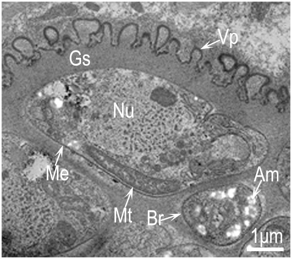
There are several reports of Sarcocystis sarcocysts in muscles of dogs, but these species have not been named. Additionally, there are two reports of Sarcocystis neurona in dogs. Here, we propose two new names, Sarcocystis caninum, and Sarcocystis svanai for sarcocysts associated with clinical muscular sarcocystosis in four domestic dogs (Canis familiaris), one each from Montana and Colorado in the USA, and two from British Columbia, Canada. Only the sarcocyst stage was identified. Most of the sarcocysts identified were S. caninum. Sarcocysts were studied using light microscopy, transmission electron microscopy (TEM), and polymerase chain reaction. Based on collective results two new species, S. caninum and S. svanai were designated. Sarcocystis caninum and S. svanai were structurally distinct. Sarcocystis caninum sarcocysts were up to 1.2 mm long and up to 75 μm wide. By light microscopy, the sarcocyst wall was relatively thin and smooth. By TEM, the sarcocyst wall was "type 9", 1-2 μm thick, and contained villar protrusions that lacked microtubules. Bradyzoites in sections were 7-9 μm long. Sarcocysts of S. svanai were few and were identified by TEM. Sarcocystis svanai sarcocysts were "type 1", thin walled (< 0.5 μm), and the wall lacked villar protrusions but had tiny blebs that did not invaginate. DNA was extracted either from infected frozen muscle biopsies or formalin-fixed paraffin-embedded sections. Dogs were either singly infected with S. caninum or multiply co-infected with S. caninum and S. svanai (the result of a mixed infection) based on multilocus DNA sequencing and morphology. BLASTn analysis established that the sarcocysts identified in these dogs were similar to, but not identical to Sarcocystis canis or Sarcocystis arctosi, parasites found to infect polar bears (Ursus maritimus) or brown bears (Ursus arctosi), respectively. However, the S. caninum sequence showed 100% identify over the 18S rRNA region sequenced to that of S. arctica, a parasite known to infect Arctic foxes (Vulpes lagopus).

Keywords: Canada; USA; dog.


Figures

Figure 1
Figure 1
Maximum likelihood SSU 18S rRNA sequence phylogenetic tree. An alignment of partial SSU 18S rRNA sequences were used to construct a phylogenetic tree to root the dog Sarcocsytis within the coccidia, and more specifically, within the Family Sarcocystidae. Reference 18S rRNA gene sequences were retrieved from the NCBI Genbank database and aligned using ClustalX. To construct the maximum likelihood phylogenetic tree, aligned sequences were directly incorporated into MEGA. The percentage of replicate trees in which the various taxa clustered together in the bootstrap test (10,000 replicates) is shown next to the branches.
Figure 2
Figure 2
Maximum likelihood phylogenetic tree obtained using the rpoB genetic marker. An alignment of the rpoB DNA sequences was used to construct phylogenetic trees to root the Sarcocystis caninum and Sarcocystis svanai sequences within the Sarcocystidae. The percentage of replicate trees in which the associated taxa clustered together in the bootstrap test (10,000 replicates) is shown next to the branches. Only nodes with greater than 50% support are resolved. The rpoB locus differentiated S. caninum from S. svanai into 2 different species that are closely related to S. canis and S. campestris, respectively.
Figure 3
Figure 3
Light microscopic structure of sarcocysts of Sarcocystis caninum, n.sp. from the naturally–infected dog B. A Low power view of sarcocysts (arrow) in triceps. Hematoxylin and eosin stain. B Higher magnification of the sarcocyst wall (indicated by opposing arrowheads) continued in to the interior of the sarcocyst as septa (S). The cyst wall appears without projections. Hematoxylin and eosin stain. C Higher magnification of the cyst wall (indicated by opposing arrowheads) in 1 μm section stained with toluidine blue. Note minute protrusions on the cyst wall, faintly stained metrocytes (Me) and darkly stained bradyzoites in longitudinal section (marked by opposing arrowheads).
Figure 4
Figure 4
TEM of an immature sarcocyst of Sarcocystis caninum, n. sp. Dog D. Note pleomorphic villar protrusion (Vp), smooth ground substance (Gs), a metrocyte (Me) with a nucleus (Nu), and cross section of a bradyzoite (Br) with numerous amylopectin granules (Am).
Figure 5
Figure 5
TEM of Sarcocystis caninum, n. sp. sarcocyst in dog B. Note cytolysis of the host cell and the presence of numerous host cell mitochondriion (Hmc) surrounding the cyst wall. The villar protrusions (Vp) are pleomorphic. The ground substance (Gs) is smooth and continued in the interior as septa (Se). Also note metrocytes (Me) with few organelles without amylopectin granules and bradyzoites (Br) with numerous micronemes, and amylopectin granules.
Figure 6
Figure 6
Higher magnification of the cyst wall of Sarcocystis caninum, n. sp. sarcocyst in dog B. Note convoluted parasitophorous vacuolar membrane (Pvm) with prominent villar protusions (Vp) that are lined by an electron dense layer (Edl), except at invaginations (arrowheads) at irregular distances. The ground substance (Gs) is smooth and juxtaposed with double layered pellicle (black arrows) of the bradyzoite.
Figure 7
Figure 7
TEM of 2 longitudinally cut bradyzoites (A, B) from 2 sarcocysts of Sarcocystis caninum, n. sp. in dog B. Note double layered pellicle, slightly thickened at the conoidal end, conoid (Co), few rhoptries (Ro), a mitrochondriion (Mt), numerous micronemes (Mn), subterminal nucleus (Nu), and numerous amylopectin granules (Am).
Figure 8
Figure 8
TEM of Sarcocystis svanai, n. sp. from dogs. (A) Note an immature sarcocyst with few metrocytes (Me), thin cyst wall with blebs (arrows), and host cell (Hc). Dog C. (B) Nearly mature sarcocyst from dog B. Note 2 bradyzoites juxtaposed with the cyst wall. The parasitophorous vacuolar membrane (Pvm) has tiny blebs on the cyst wall. The ground substance (Gs) is smooth. Note conoid (Co), rhoptries (Ro), numerous micronemes (Mi), and amylopectin granules (Am). (C) Details of the sarcocyst wall shown in 8A. Note wavy parasitophorous vacuolar membrane (Pvm) with blebs, and smooth ground substance (Gs). Also note pellicle (black arrows) of the bradyzoite (Br) juxtaposed with Gs. Also note host cell mitochondrion (Hmc).
Figure 9
Figure 9
TEM of a sarcocyst of Sarcocystis svanai, n. sp. from dog B. Note thin cyst wall with a smooth ground substance (Gs), and wavy parasitophorous vacuolar membrane (Pvm). Note a maturing bradyzoite (Br) with a conoid (Co), nucleus (Nu), and amylopectin granules (Am).
Figure 10
Figure 10
Comparison of the sarcocyst walls of 2 species of Sarcocystis. A Sarcocystis caninum, n. sp. B Sarcocystis svanai, n. sp. The cyst wall (Cw) of S. caninum is more than twice thicker than that of S. svanai. Also note, for orientation, the host cell (Hc), host cell mitrochondriion (Hmc), villar protrusions (Vp), ground substance layer (Gs) juxtaposed to bradyzoite (Br), and double layered pellicle (black arrows).

References

    1. Blagburn BL, Braund KG, Amling KA, Toivio-Kinnucan M. Muscular Sarcocystis in a dog. Proc Helminthol Soc Wash. 1989;56:207–210.
    1. Britton AP, Dubey JP, Rosenthal BM. Rhinitis and disseminated disease in a ferret (Mustela putorius furo) naturally infected with Sarcocystis neurona. Vet Parasitol. 2010;169:226–231. - PubMed
    1. Bwangamoi O, Ngatia TA, Richardson JD. Sarcocystis-like organisms in musculature of a domestic dog (Canis familiaris) and wild dogs (Lycaon pictus) in Kenya. Vet Parasitol. 1993;49:201–205. - PubMed
    1. Carlson-Bremer D, Johnson CK, Miller RH, Gulland FMD, Conrad PA, Wasmuth JD, Colegrove KM, Grigg ME. Identification of two novel coccidian species shed by California sea lions (Zalophus californianus) J Parasitol. 2012;98:347–354. - PMC - PubMed
    1. Cawthorn RJ, Wobeser GA, Gajadhar AA. Description of Sarcocystis campestris sp n (Protozoa: Sarcocystidae): a parasite of the badger Taxidea taxus with experimental transmission to the Richardson’s ground squirrel, Spermophilus richardsonii. Can J Zool. 1983;61:370–377.

Publication types

MeSH terms

Associated data