

Dissection of glucocorticoid receptor-mediated inhibition of the hypothalamic-pituitary-adrenal axis by gene targeting in mice
- PMID: 25256348
- PMCID: PMC4342273
- DOI: 10.1016/j.yfrne.2014.09.002
Dissection of glucocorticoid receptor-mediated inhibition of the hypothalamic-pituitary-adrenal axis by gene targeting in mice
Abstract
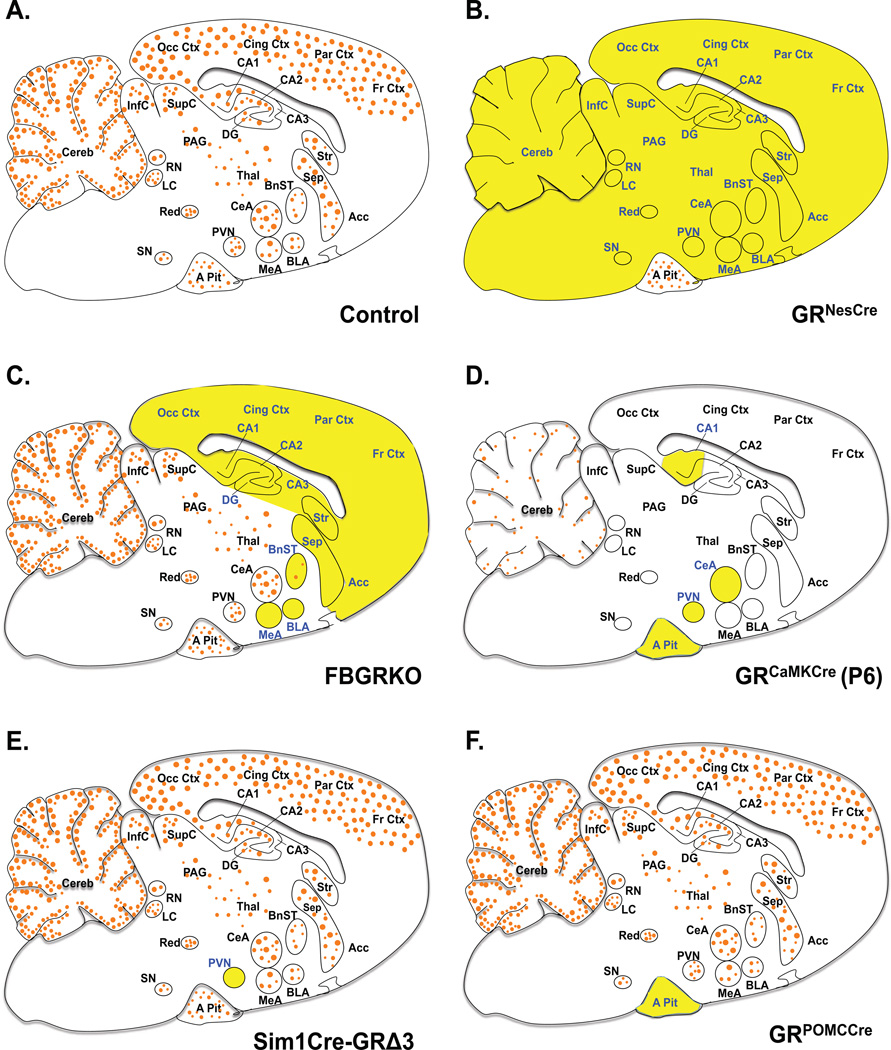
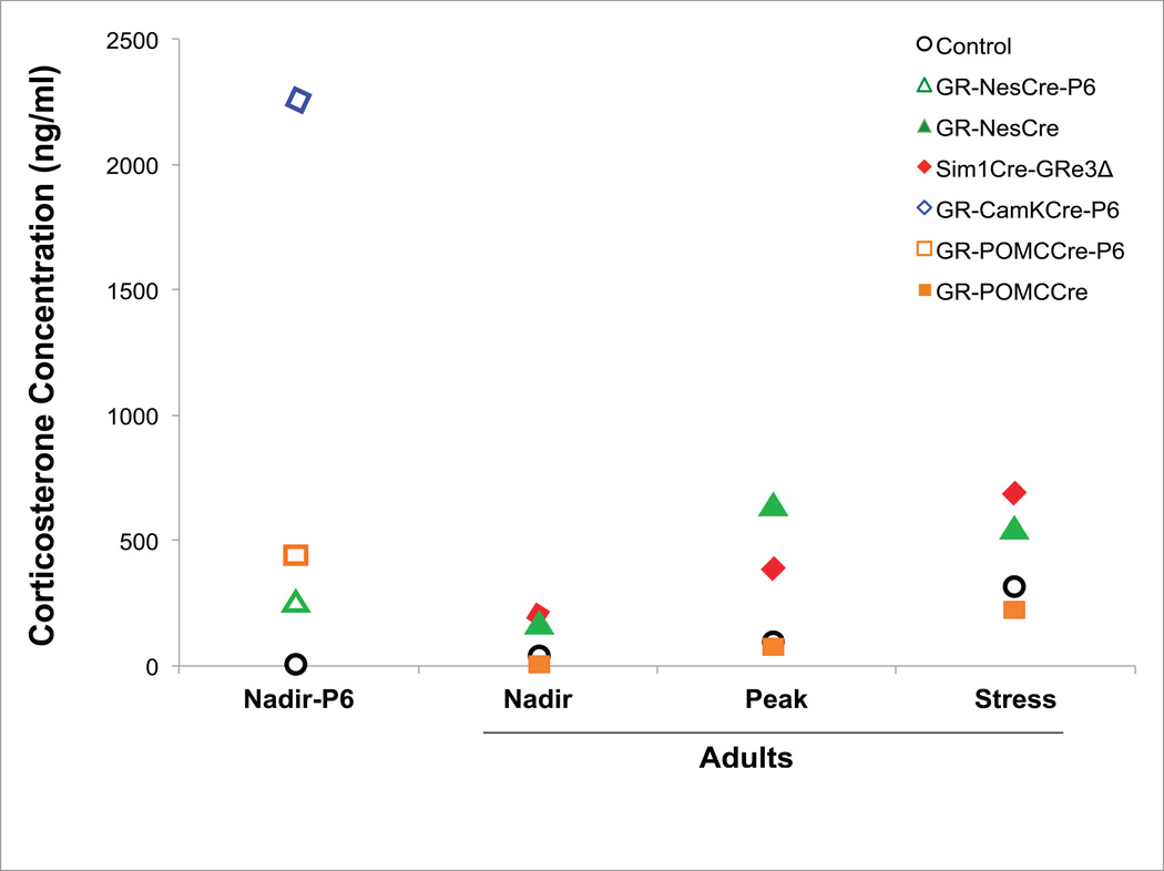
Negative feedback regulation of glucocorticoid (GC) synthesis and secretion occurs through the function of glucocorticoid receptor (GR) at sites in the hypothalamic-pituitary-adrenal (HPA) axis, as well as in brain regions such as the hippocampus, prefrontal cortex, and sympathetic nervous system. This function of GRs in negative feedback coordinates basal glucocorticoid secretion and stress-induced increases in secretion that integrate GC production with the magnitude and duration of the stressor. This review describes the effects of GR loss along major sites of negative feedback including the entire brain, the paraventricular nucleus of the hypothalamus (PVN), and the pituitary. In genetic mouse models, we evaluate circadian regulation of the HPA axis, stress-stimulated neuroendocrine response and behavioral activity, as well as the integrated response of organism metabolism. Our analysis provides information on contributions of region-specific GR-mediated negative feedback to provide insight in understanding HPA axis dysregulation and the pathogenesis of psychiatric and metabolic disorders.
Keywords: Adrenocorticotropic hormone; Circadian regulation; Corticosterone; Corticotropin-releasing hormone; Glucocorticoid receptor; Hypothalamic–pituitary–adrenal axis; Metabolism; Paraventricular nucleus of the hypothalamus; Pituitary; Stress.
Copyright © 2014 Elsevier Inc. All rights reserved.
Figures
References
-
- Ambroggi F, Turiault M, Milet A, Deroche-Gamonet V, Parnaudeau S, Balado E, Barik J, van der Veen R, Maroteaux G, Lemberger T, Schütz G, Lazar M, Marinelli M, Piazza PV, Tronche F. Stress and addiction: glucocorticoid receptor in dopaminoceptive neurons facilitates cocaine seeking. Nat. Neurosci. 2009;12:247–249. - PubMed
-
- Atalar F, Gormez S, Caynak B, Akan G, Tanriverdi G, Bilgic-Gazioglu S, Gunay D, Duran C, Akpinar B, Ozbek U, Buyukdevrim AS, Yazici Z. The role of mediastinal adipose tissue 11β-hydroxysteroid d ehydrogenase type 1 and glucocorticoid expression in the development of coronary atherosclerosis in obese patients with ischemic heart disease. Cardiovasc. Diabetol. 2012;11:115. - PMC - PubMed
-
- Balsalobre A, Brown SA, Marcacci L, Tronche F, Kellendonk C, Reichardt HM, Schütz G, Schibler U. Resetting of circadian time in peripheral tissues by glucocorticoid signaling. Science. 2000;289:2344–2347. - PubMed
-
- Balthasar N, Dalgaard LT, Lee CE, Yu J, Funahashi H, Williams T, Ferreira M, Tang V, McGovern RA, Kenny CD, Christiansen LM, Edelstein E, Choi B, Boss O, Aschkenasi C, Zhang C, Mountjoy K, Kishi T, Elmquist JK, Lowell BB. Divergence of melanocortin pathways in the control of food intake and energy expenditure. Cell. 2005;123:493–505. - PubMed
Publication types
MeSH terms
Substances
Grants and funding
LinkOut - more resources
Full Text Sources
Other Literature Sources
Miscellaneous
