. 2014 Sep 26:11:167.
doi: 10.1186/s12974-014-0167-6.

Neuroprotective effects of bilobalide on cerebral ischemia and reperfusion injury are associated with inhibition of pro-inflammatory mediator production and down-regulation of JNK1/2 and p38 MAPK activation

Neuroprotective effects of bilobalide on cerebral ischemia and reperfusion injury are associated with inhibition of pro-inflammatory mediator production and down-regulation of JNK1/2 and p38 MAPK activation

Mingjin Jiang et al. J Neuroinflammation. .

Abstract

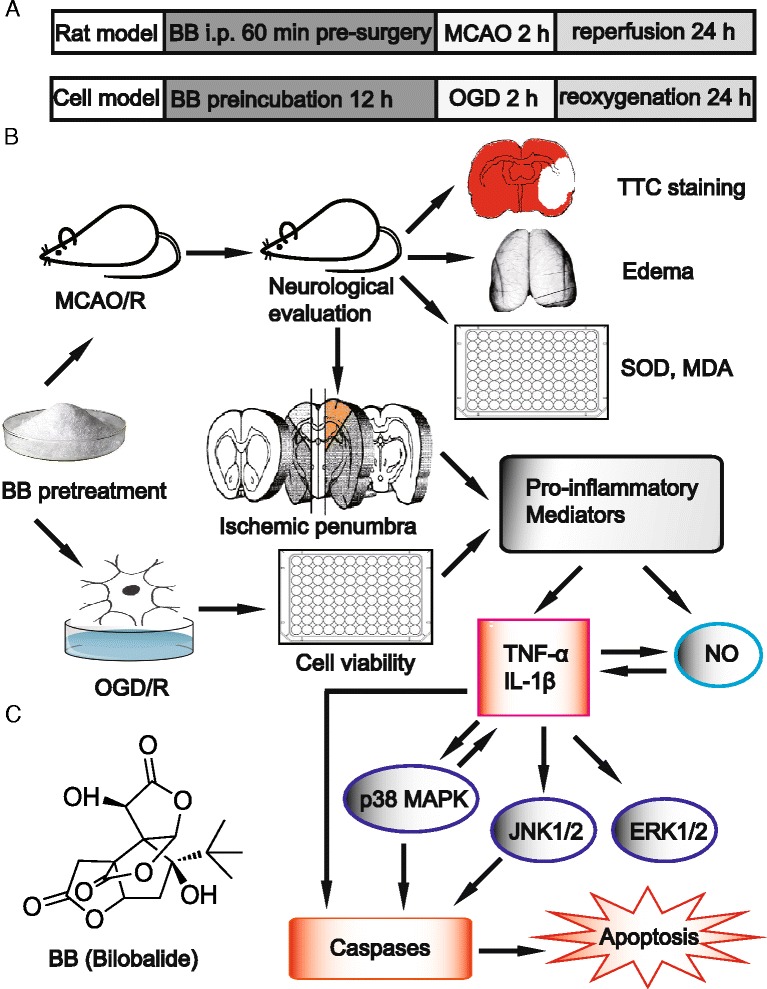
Background: Mitogen-activated protein kinase (MAPK) signaling pathways are implicated in inflammatory and apoptotic processes of cerebral ischemia and reperfusion (I/R) injury. Hence, MAPK pathways represent a promising therapeutic target. Exploring the full potential of inhibitors of MAPK pathways is a useful therapeutic strategy for ischemic stroke. Bilobalide, a predominant sesquiterpene trilactone constituent of Ginkgo biloba leaves, has been shown to exert powerful neuroprotective properties, which are closely related to both anti-inflammatory and anti-apoptotic pathways. We investigated the neuroprotective roles of bilobalide in the models of middle cerebral artery occlusion and reperfusion (MCAO/R) and oxygen-glucose deprivation and reoxygenation (OGD/R) of cerebral I/R injury. Moreover, we attempted to confirm the hypothesis that its protection effect is via modulation of pro-inflammatory mediators and MAPK pathways.

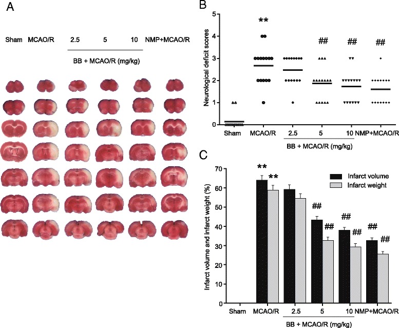
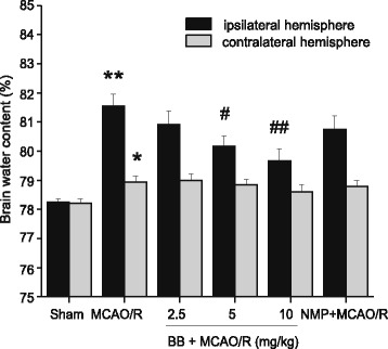
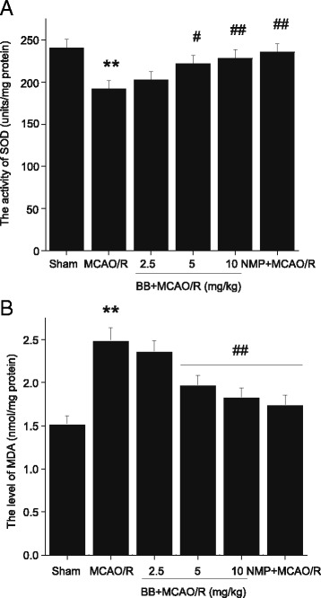
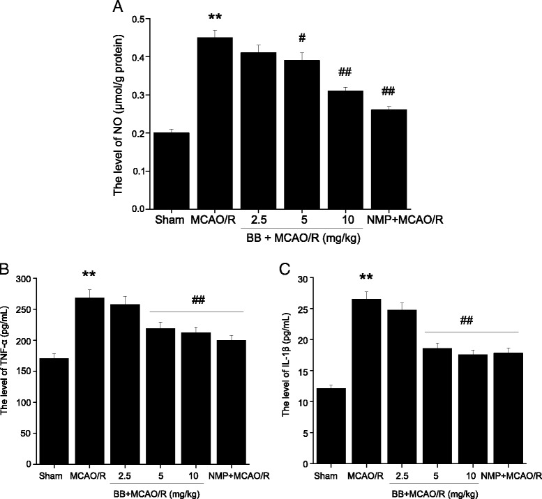
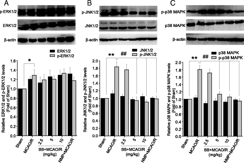
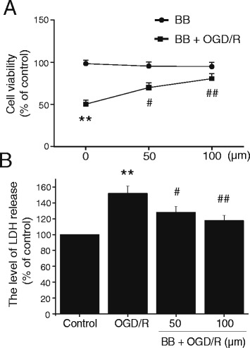
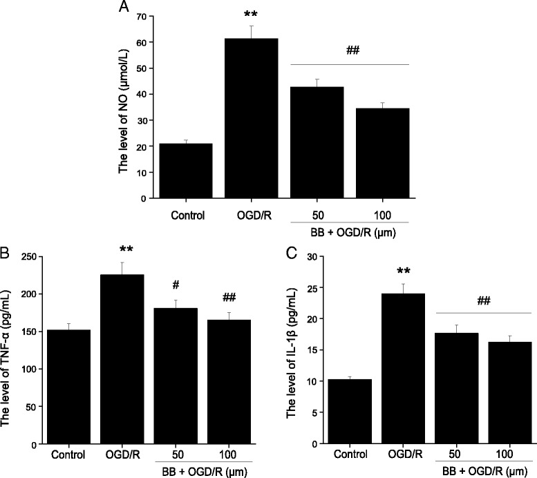
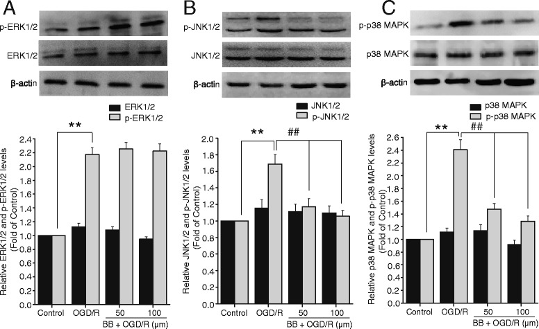
Methods: Male Sprague-Dawley rats were subjected to MCAO for 2 h followed by reperfusion for 24 h. Bilobalide was administered intraperitoneally 60 min before induction of middle cerebral artery occlusion (MCAO). After reperfusion, neurological deficit scores, infarct volume, infarct weight, and brain edema were assessed. Ischemic penumbrae of the cerebral cortex were harvested to determine superoxide dismutase (SOD), malondialdehyde (MDA), nitric oxide, TNF-α, interleukin 1β (IL-1β), p-ERK1/2, p-JNK1/2, and p-p38 MAPK concentration. Similarly, the influence of bilobalide on the expression of nitric oxide, TNF-α, IL-1β, p-ERK1/2, p-JNK1/2, and p-p38 MAPK was also observed in an OGD/R in vitro model of I/R injury.

Results: Pretreatment with bilobalide (5, 10 mg/kg) significantly decreased neurological deficit scores, infarct volume, infarct weight, brain edema, and concentrations of MDA, nitric oxide, TNF-α, IL-1β, and increased SOD activity. Furthermore, bilobalide (5, 10 mg/kg) pretreatment significantly down-regulated both p-JNK1/2 and p-p38 MAPK expression, whereas they had no effect on p-ERK1/2 expression in the ischemic penumbra. Supporting these observations in vivo, pretreatment with bilobalide (50, 100 μM) significantly down-regulated nitric oxide, TNF-α, IL-1β, p-JNK1/2, and p-p38 MAPK expression, but did not change p-ERK1/2 expression in rat cortical neurons after OGD/R injury.

Conclusions: These data indicate that the neuroprotective effects of bilobalide on cerebral I/R injury are associated with its inhibition of pro-inflammatory mediator production and down-regulation of JNK1/2 and p38 MAPK activation.


Figures

Figure 1
Figure 1
Chemical structure of bilobalide and experimental protocol. (A) Rat MCAO/R and in vitro OGD/R model of cerebral ischemia and reperfusion injury. Bilobalide (2.5, 5, and 10 mg/kg) was administered via a single intraperitoneal injection 60 min prior to surgery in the rat model. In the cell model, cortical neurons were previously cultured in bilobalide (50, 100 μM) for 12 h by dissolving bilobalide in serum-free DMEM. (B) Experimental protocol; neuroprotective effects of bilobalide on cerebral ischemia and reperfusion injury are associated with pro-inflammatory mediator production and MAPK signaling pathway. (C) Chemical structure of bilobalide. BB, bilobalide; MCAO/R, middle cerebral artery occlusion and reperfusion; MDA, malondialdehyde; OGD/R, oxygen-glucose deprivation and reoxygenation; SOD, superoxide dismutase; TTC, 2,3,5-triphenyltetrazolium chloride.
Figure 2
Figure 2
Effects of bilobalide on neurological deficit scores, infarct volume, and infarct weight. (A) Representative photographs of brain slices following infarction, stained with 2, 3, 5-triphenyltetrazolium chloride. Red tissue is healthy; white tissue is infarcted. Pretreatment with bilobalide (5, 10 mg/kg) significantly reduced infarct size (B) and improved neurological scores (C) and decreased infarct volume and infarct weight compared with the MCAO/R group. Mean values ± standard error of the mean for 15 (neurological evaluation) and six rats per group. ** P < 0.01 versus sham; ## P < 0.01 versus MCAO/R. BB, bilobalide; MCAO/R, middle cerebral artery occlusion and reperfusion; NMP, nimodipine.
Figure 3
Figure 3
Effect of bilobalide on brain water content. Pretreatment with bilobalide (5, 10 mg/kg) significantly reduced brain water content in the ipsilateral hemisphere, but did not affect brain water content in the contralateral hemisphere compared with the MCAO/R group. Mean values ± standard error of the mean for six rats per group. * P < 0.05, ** P < 0.01 versus sham; # P < 0.05, ## P < 0.01 versus MCAO/R. BB, bilobalide; MCAO/R, middle cerebral artery occlusion and reperfusion; NMP, nimodipine.
Figure 4
Figure 4
Effects of bilobalide on the levels of SOD and MDA. (A) Pretreatment with bilobalide (5, 10 mg/kg) significantly increased SOD activity (B) and decreased MDA concentration compared with the MCAO/R group. Mean values ± standard error of the mean for six rats per group. ** P < 0.01 versus sham; # P < 0.05, ## P < 0.01 versus MCAO/R. BB, bilobalide; MCAO/R, middle cerebral artery occlusion and reperfusion; NMP, nimodipine.
Figure 5
Figure 5
Effects of bilobalide on the levels of nitric oxide, TNF-α, and IL-1β in ischemic penumbra after MCAO/R. (A) Pretreatment with bilobalide (5, 10 mg/kg) significantly reduced concentrations of nitric oxide (B) and TNF-α (C) and IL-1β in the ischemic penumbra compared with the MCAO/R group. Mean values ± standard error of the mean for six rats per group. ** P < 0.01 versus sham; # P < 0.05, ## P < 0.01 versus MCAO/R. BB, bilobalide; MCAO/R, middle cerebral artery occlusion and reperfusion; NMP, nimodipine.
Figure 6
Figure 6
Effects of bilobalide on the expression of ERK1/2, JNK1/2, and p38 MAPK in ischemic penumbra after MCAO/R. (A) Pretreatment with bilobalide (5, 10 mg/kg) did not change p-ERK1/2 concentration, (B) significantly reduced p-JNK1/2 concentration, (C) and remarkably decreased p-p38 MAPK concentration in the ischemic penumbra compared with the MCAO/R group. There were no obvious differences in the levels of total ERK1/2, total JNK1/2, and total p38 MAPK among all experimental groups. Mean values ± standard error of the mean for six rats per group. * P < 0.05, ** P < 0.01 versus sham; ## P < 0.01 versus MCAO/R. BB, bilobalide; MCAO/R, middle cerebral artery occlusion and reperfusion; NMP, nimodipine.
Figure 7
Figure 7
Effects of bilobalide on cell viability in primary cortical neurons after OGD/R. (A) Incubation of cortical neurons with different concentrations of bilobalide (50, 100 μM) alone for 12 h did not affect cell viability. After cortical neurons were exposed to 2 h of OGD following 24 h of reoxygenation, cell viability was significantly decreased; pretreatment with bilobalide (50, 100 μM) for 12 h significantly the cell viability. (B) After cortical neurons were exposed to 2 h of OGD following 24 h of reoxygenation, the level of LDH release was remarkably increased; pretreatment with bilobalide (50, 100 μM) for 12 h significantly decreased the LDH level. Mean values ± standard error of the mean ** P < 0.01 versus control; # P < 0.05, ## P < 0.01 versus. OGD/R. BB, bilobalide; LDH, lactate dehydrogenase; OGD/R, oxygen-glucose deprivation and reoxygenation.
Figure 8
Figure 8
Effects of bilobalide on the levels of nitric oxide, TNF-α, and IL-1β in primary cortical neurons after OGD/R. (A) Pretreatment with bilobalide (50, 100 μM) significantly reduced nitric oxide (B) and TNF-α (C) and IL-1β levels in OGD/R-induced cortical neurons compared with the OGD/R group. Mean values ± standard error of the mean ** P < 0.01 versus control; # P < 0.05, ## P < 0.01 versus OGD/R. BB, bilobalide; OGD/R, oxygen-glucose deprivation and reoxygenation.
Figure 9
Figure 9
Effects of bilobalide on the expression of ERK1/2, JNK1/2, and p38 MAPK in primary cortical neurons after OGD/R. (A) Pretreatment with bilobalide (50, 100 μM) did not have an effect on p-ERK1/2 level (B) and significantly reduced p-JNK1/2 level (C) and remarkably down-regulated p-p38 MAPK expression in OGD/R-induced cortical neurons compared with the OGD/R group. No significant differences were observed in the expression of total ERK1/2, total JNK1/2, and total p38 MAPK among all experimental groups in vitro. Mean values ± standard error of the mean ** P < 0.01 versus control; ## P < 0.01 versus OGD/R. BB, bilobalide; OGD/R, oxygen-glucose deprivation and reoxygenation.

References

    1. Go AS, Mozaffarian D, Roger VL, Benjamin EJ, Berry JD, Blaha MJ, Dai S, Ford ES, Fox CS, Franco S, Fullerton HJ, Gillespie C, Hailpern SM, Heit JA, Howard VJ, Huffman MD, Judd SE, Kissela BM, Kittner SJ, Lackland DT, Lichtman JH, Lisabeth LD, Mackey RH, Magid DJ, Marcus GM, Marelli A, Matchar DB, McGuire DK, Mohler ER, 3rd, Moy CS, et al. Heart disease and stroke statistics - 2014 update: a report from the American Heart Association. Circulation. 2014;129:e28–e292. doi: 10.1161/01.cir.0000441139.02102.80. - DOI - PMC - PubMed
    1. Jauch EC, Saver JL, Adams HP, Jr, Bruno A, Connors JJ, Demaerschalk BM, Khatri P, McMullan PW, Jr, Qureshi AI, Rosenfield K, Scott PA, Summers DR, Wang DZ, Wintermark M, Yonas H, American Heart Association Stroke Council; Council on Cardiovascular Nursing; Council on Peripheral Vascular Disease; Council on Clinical Cardiology Guidelines for the early management of patients with acute ischemic stroke: a guideline for healthcare professionals from the American Heart Association/American Stroke Association. Stroke. 2013;44:870–947. doi: 10.1161/STR.0b013e318284056a. - DOI - PubMed
    1. Eltzschig HK, Eckle T. Ischemia and reperfusion - from mechanism to translation. Nat Med. 2011;17:1391–1401. doi: 10.1038/nm.2507. - DOI - PMC - PubMed
    1. Iadecola C, Alexander M. Cerebral ischemia and inflammation. Curr Opin Neurol. 2001;14:89–94. doi: 10.1097/00019052-200102000-00014. - DOI - PubMed
    1. Broughton BR, Reutens DC, Sobey CG. Apoptotic mechanisms after cerebral ischemia. Stroke. 2009;40:e331–e339. doi: 10.1161/STROKEAHA.108.531632. - DOI - PubMed

Publication types