Review
. 2014 Oct;7(10):1131-42.
doi: 10.1242/dmm.016428.

Microbial imbalance and intestinal pathologies: connections and contributions

Affiliations
Review

Microbial imbalance and intestinal pathologies: connections and contributions

Ye Yang et al. Dis Model Mech. 2014 Oct.

Abstract

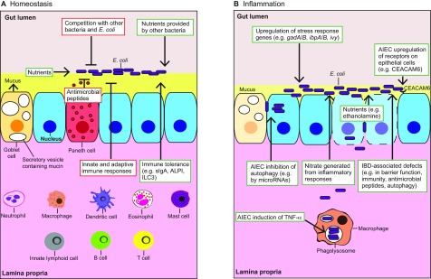
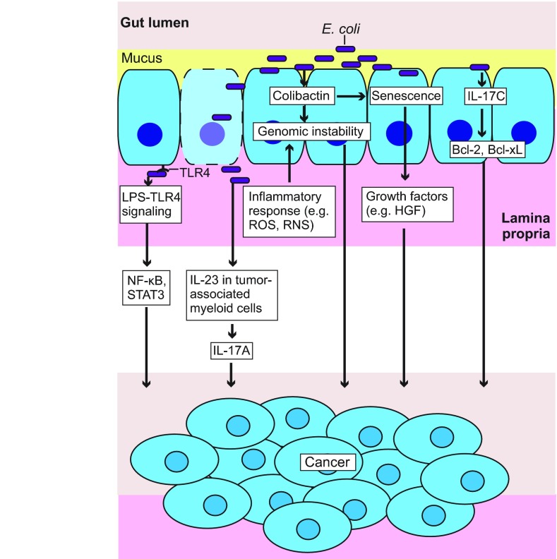
Microbiome analysis has identified a state of microbial imbalance (dysbiosis) in patients with chronic intestinal inflammation and colorectal cancer. The bacterial phylum Proteobacteria is often overrepresented in these individuals, with Escherichia coli being the most prevalent species. It is clear that a complex interplay between the host, bacteria and bacterial genes is implicated in the development of these intestinal diseases. Understanding the basic elements of these interactions could have important implications for disease detection and management. Recent studies have revealed that E. coli utilizes a complex arsenal of virulence factors to colonize and persist in the intestine. Some of these virulence factors, such as the genotoxin colibactin, were found to promote colorectal cancer in experimental models. In this Review, we summarize key features of the dysbiotic states associated with chronic intestinal inflammation and colorectal cancer, and discuss how the dysregulated interplay between host and bacteria could favor the emergence of E. coli with pathological traits implicated in these pathologies.

Keywords: Adherent-invasive E. coli; CRC; Colibactin; Dysbiosis; IBD.


Figures

Fig. 1.
Fig. 1.
Events modulating E. coli colonization and fitness in the intestine. Events promoting E. coli fitness are illustrated in green boxes and those inhibiting E. coli growth are shown in red boxes. (A) At homeostasis, bacteria such as E. coli are prevented from attaching to epithelial cells (blue cells; for simplicity, villi are not shown) by a mucus layer present at the apical surface of the gut epithelium. The host immune system and the Paneth-cell-derived antimicrobial peptides regulate microbial growth. Major cell types that are involved in immune regulation of the gut microbes are illustrated in the lamina propria, where they are found. The host gut provides nutrients to support E. coli colonization, and immune tolerance mechanisms help to maintain a healthy E. coli community in the intestine. E. coli colonization is also modulated by bacterial competition for nutrients. Other bacteria can provide nutrients (e.g. mono- and disaccharides) to E. coli and promote its growth. (B) During intestinal inflammation, mucus depletion facilitates E. coli attachment to the epithelium. E. coli adjust their metabolism, and upregulate stress response genes to promote their survival. E. coli can take advantage of inflammation by using nutrients provided by dead cells and by respiring host-derived nitrate. Adherent invasive E. coli (AIEC) can disrupt the epithelial barrier, inhibit epithelial cell autophagy to promote intracellular survival, and upregulate surface receptors on epithelial cells to increase adherence. AIEC infection stimulates TNF-α expression in macrophages, which promotes their intracellular replication. IBD-associated deficiencies such as reduced secretion of antimicrobial peptides due to Paneth cell dysfunction (Paneth cells not shown) can further allow bacterial attachment, invasion and growth. CEACAM6, carcinoembryonic antigen-related cell adhesion molecule 6; gadA/B, glutamic acid decarboxylase A and B; ALPI, intestinal alkaline phosphatase; ibpA/B, inclusion body protein A and B; ILC3, group 3 innate lymphoid cells; ivy, inhibitor of C-type lysozyme; sIgA, secretory immunoglobulin A; TNF-α, tumor necrosis factor α.
Fig. 2.
Fig. 2.
Intestinal inflammation creates a different environment for the gut microbes. Hematoxylin and eosin (H&E)-stained colon Swiss roll sections from a 9-month-old wild-type (WT) mouse (left; healthy) and an age-matched interleukin-10 knockout (Il-10−/−) mouse (right, inflamed). (Left, inset) The WT mouse colon has a well-organized lining of villi (gray box), with the presence of columnar epithelial cells (with microvilli; blue arrow) and goblet cells (black arrow). (Right, inset) The inflamed Il-10−/− mouse colon shows loss of the regular villus structures, thickening of the mucosa, crypt elongation (brown arrow), infiltration of inflammatory immune cells (yellow arrow), depletion of goblet cells, disruption of the brush border (red arrow) and loss of surface epithelial cells (green arrow).
Fig. 3.
Fig. 3.
E. coli cancer-promoting mechanisms. E. coli-induced innate immune responses (e.g. LPS-TLR4 signaling) can result in enhanced cell proliferation and survival through activating NF-κB and STAT3. Bacterial products (e.g. LPS) can promote cell proliferation by activating myeloid-cell-derived IL-23 and subsequently IL-17A. E. coli can induce epithelial-cell-derived IL-17C and subsequently Bcl-2 and Bcl-xL to enhance cell survival. Reactive oxygen and nitrogen species (ROS, RNS) produced by innate immune cells (e.g. macrophages and neutrophils) during E. coli infection, and E. coli-derived genotoxins (e.g. colibactin) can cause DNA damage and thus genomic instability. Colibactin-producing E. coli infection causes epithelial cell senescence, and senescent cells release growth factors (e.g. HGF) to promote cell proliferation. Bcl-2, B-cell lymphoma 2; Bcl-xL, B-cell lymphoma-extra large; HGF, hepatocyte growth factor; IL-17A, interleukin 17A; IL-17C, interleukin 17C; IL-23, interleukin 23; LPS, lipopolysaccharide; NF-κB (nuclear factor kappa-light-chain-enhancer of activated B cells; ROS, reactive oxygen species; RNS, reactive nitrogen species; STAT3, signal transducer and activator of transcription 3; TLR4, Toll-like receptor 4.

References

    1. Adolph T. E., Tomczak M. F., Niederreiter L., Ko H. J., Böck J., Martinez-Naves E., Glickman J. N., Tschurtschenthaler M., Hartwig J., Hosomi S., et al. (2013). Paneth cells as a site of origin for intestinal inflammation. Nature 503, 272–276 - PMC - PubMed
    1. Ahn J., Sinha R., Pei Z., Dominianni C., Wu J., Shi J., Goedert J. J., Hayes R. B., Yang L. (2013). Human gut microbiome and risk for colorectal cancer. J. Natl. Cancer Inst. 105, 1907–1911 - PMC - PubMed
    1. Alpert C., Scheel J., Engst W., Loh G., Blaut M. (2009). Adaptation of protein expression by Escherichia coli in the gastrointestinal tract of gnotobiotic mice. Environ. Microbiol. 11, 751–761 - PubMed
    1. Apperloo-Renkema H. Z., Van der Waaij B. D., Van der Waaij D. (1990). Determination of colonization resistance of the digestive tract by biotyping of Enterobacteriaceae. Epidemiol. Infect. 105, 355–361 - PMC - PubMed
    1. Arpaia N., Campbell C., Fan X., Dikiy S., van der Veeken J., deRoos P., Liu H., Cross J. R., Pfeffer K., Coffer P. J., et al. (2013). Metabolites produced by commensal bacteria promote peripheral regulatory T-cell generation. Nature 504, 451–455 - PMC - PubMed

Publication types

MeSH terms