Review
. 2014 Sep 26:2:142.
doi: 10.1186/s40478-014-0142-6.

Microglial pathology

Review

Microglial pathology

Wolfgang J Streit et al. Acta Neuropathol Commun. .

Abstract

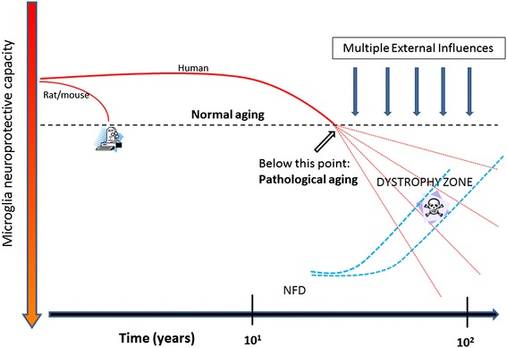
This paper summarizes pathological changes that affect microglial cells in the human brain during aging and in aging-related neurodegenerative diseases, primarily Alzheimer's disease (AD). It also provides examples of microglial changes that have been observed in laboratory animals during aging and in some experimentally induced lesions and disease models. Dissimilarities and similarities between humans and rodents are discussed in an attempt to generate a current understanding of microglial pathology and its significance during aging and in the pathogenesis of Alzheimer dementia (AD). The identification of dystrophic (senescent) microglia has created an ostensible conflict with prior work claiming a role for activated microglia and neuroinflammation during normal aging and in AD, and this has raised a basic question: does the brain's immune system become hyperactive (inflamed) or does it become weakened (senescent) in elderly and demented people, and what is the impact on neuronal function and cognition? Here we strive to reconcile these seemingly contradictory notions by arguing that both low-grade neuroinflammation and microglial senescence are the result of aging-associated free radical injury. Both processes are damaging for microglia as they synergistically exhaust this essential cell population to the point where the brain's immune system is effete and unable to support neuronal function.


Figures

Figure 1
Figure 1
Microglial senescence may be a key event in aging and Alzheimer’s disease. Free radical injury occurs naturally during aging affecting both microglia and neurons. It contributes to microglial senescence directly by causing oxidative injury in microglia, and indirectly by causing oxidative injury in neurons which elicits a low-level inflammatory response. The latter causes chronic stress to microglia contributing to exhaustion and senescence. If normal aging is exacerbated by acute injuries eliciting intense inflammation, this may accelerate naturally occurring microglial senescence.
Figure 2
Figure 2
Dystrophic microglia have many guises and can be stained with different antibodies. A, B Iba1 (red) and CD68 (green) double immunohistochemical staining of dystrophic microglia in AD brain, as seen by 3-D confocal microscopy. Note fragmentation of cytoplasmic processes and punctate CD68 labeling indicating intracellular location of lysosomal antigen. C-E, anti-HLA-DR antigen immunohistochemistry (LN-3 antibody) shows dystrophic spheroid formation (C), atrophic deramified processes (D), and formation of microglial aggregate with spheroids (E). F, ferritin-positive fragmented microglia. G, Iba1-positive dystrophic microglia with beaded, fragmenting processes and spheroids. All images taken in cerebral cortex of AD subjects. Scale bars: 10 μm (A, B), 20 μm (C, D, G), 40 μm (E, F).
Figure 3
Figure 3
Schematic representation of cytorrhexis development. Ramified cells (1) develop spheroidal swellings (2), which progress to beading and partial fragmentation of processes (3), which progresses to complete fragmentation while still maintaining cell contours (4), eventually ending up as scattered fragments (5). Note that the cell nucleus remains intact throughout.
Figure 4
Figure 4
Different functional states of microglia can be defined morphologically. A, ramified microglia exhibit highly branched processes with which they explore their surrounding microenvironment. B, activated microglia retract processes and become enlarged due to organelle build-up and increased metabolic activity. C, phagocytic microglia often appear as rounded brain macrophages. D, dystrophic microglia most characteristically display beaded, twisted or fragmented processes. Human cerebral cortex stained with Iba1 antibody. Bar = 20 μm.
Figure 5
Figure 5
Histopathological differences in microglial aging between laboratory rodents and humans provide a foundation for normal and pathological aging. Mice and rats experience only normal aging due to their short life spans and controlled environment, whereas humans who live much longer lives are exposed to diverse environmental influences and lifestyles, including diets, physical and mental activities, drugs, pollutants, comorbidities and infections, all of which may affect the rates at which microglial senescence (dystrophy) occurs, indicated by different trajectories of dashed red lines. As dystrophy occurs to varying degrees there is a decline in microglial neuroprotection and with that neurofibrillary degeneration (NFD) increases at variable rates in different individuals.
Figure 6
Figure 6
Widespread dystrophy and absence of brain macrophages are evident in Down syndrome brain. Iba1-stained section of cerebral cortex from a subject with Down syndrome with Braak stage VI neurodegeneration. Normal microglial morphology is barely discernable in only few cells and the field is mostly occupied by microglial membrane fragments of varying shapes and sizes. Scale bar: 100 μm.
Figure 7
Figure 7
Absence of activated microglia near neurons with Lewy bodies. Double-label immunostaining for microglia (Iba1, brown) and α-synuclein (black) in a subject with PD, stage 5. Section of neocortex shows Lewy bodies in cortical neurons (arrows) surrounded by normal, ramified microglia.
Figure 8
Figure 8
Ramified and dystrophic microglia are associated with amyloid-beta protein deposits. Double-label immunohistochemistry for Aβ protein (brown reaction product; 4G8 antibody) and microglia (black reaction product; Iba1 antibody) in human cerebral cortex. A, diffuse Aβ deposits are colocalized with ramified, non-activated microglia, B, as Aβ deposits become more compact microglia begin to show signs of dystrophy (arrow points to early fragmentation and spheroid formation). C, D show several advanced dense-core plaques where the dense core is comprised of dystrophic microglial fragments (white arrows). Note multiple microglial fragments in the immediate vicinty of Aβ deposits. Bar = 50 μm.
Figure 9
Figure 9
Microglia in the spinal cord of SOD G93A rats. A-C, double staining for microglia (Iba1) and neurons (NeuN) shows progressive loss of neurons during symptomatic and end stages of motor neuron disease. Microglial activation during symptomatic disease (B) progresses to microglial dystrophy at 6 months of age when animals are terminal (C). Panel D shows close-up of fragmented microglia as seen with OX-42 staining (brown color) during end stage disease. A few surviving motoneurons are stained blue with cresyl violet. Scale bars: 100 μm (A-C); 20 μm (D).
Figure 10
Figure 10
Signs of microglial dysfunction. A, Langhans type giant cell consisting of fused microglia in the brainstem of a SOD1G93A rat, as shown by lectin histochemistry (brown color). Note the characteristic arrangement of nuclei along periphery. B, nidus of bacilli in another section from the same animal, possibly a reflection of impaired bactericidal activity of microglia. Cresyl violet stain. Scale bar = 20 μm.
Figure 11
Figure 11
Microglial pathology is evident in rats exposed to nerve agent. Double immunofluorescent staining for microglia (OX-42) and cytokines (MCP-1 in A; IL-1α in B) in soman exposed rats. Panel A shows severe dystrophy of microglia, evident as extensive fragmentation of the cells’ cytoplasm, in the piriform cortex (arrows). MCP-1 immunoreactivity is localized in neurons. In Panel B, OX-42-positive microglia with fragmented processes also reveal IL-1α immunoreactivity in their cytoplasm (arrow). These cells showing features of both activation (IL-1) and degeneration (cytorrhexis) are thought to represent transitional forms; they were found in the dentate gyrus. Survival time is 12 hours; DAPI counterstain. Scale bar: 50 μm. Images provided by Erik A. Johnson, US Army Medical Research Institute of Chemical Defense (USAMRICD), Aberdeen Proving Ground, MD.

References

    1. v Eitzen U, Egensperger R, Kosel S, Grasbon-Frodl EM, Imai Y, Bise K, Kohsaka S, Mehraein P, Graeber MB. Microglia and the development of spongiform change in Creutzfeldt-Jakob disease. J Neuropathol Exp Neurol. 1998;57(3):246–256. - PubMed
    1. Mosher KI, Wyss-Coray T. Microglial dysfunction in brain aging and Alzheimer’s disease. Biochem Pharmacol. 2014;88(4):594–604. - PMC - PubMed
    1. Prokop S, Miller KR, Heppner FL. Microglia actions in Alzheimer’s disease. Acta Neuropathol. 2013;126(4):461–477. - PubMed
    1. Aguzzi A, Barres BA, Bennett ML. Microglia: scapegoat, saboteur, or something else? Science. 2013;339(6116):156–161. - PMC - PubMed
    1. Biber K, Owens T, Boddeke E. What is microglia neurotoxicity (Not)? Glia. 2014;62(6):841–854. - PubMed

Publication types