

Comment
doi: 10.1038/cr.2014.124.
Epub 2014 Sep 26.
Large-field high-resolution two-photon digital scanned light-sheet microscopy
Affiliations
- PMID: 25257466
- PMCID: PMC4650563
- DOI: 10.1038/cr.2014.124
Item in Clipboard
Comment
Large-field high-resolution two-photon digital scanned light-sheet microscopy
Cell Res.
2015 Feb.
No abstract available
Figures
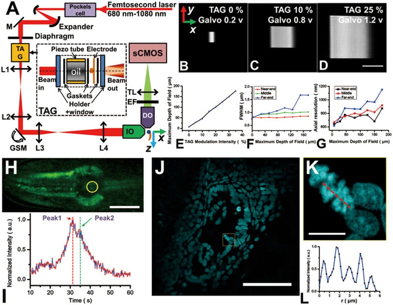
(A) A schematic illustration of 2P3A-DSLM. We used a commercial Ti-sapphire femtosecond laser (Chameleon Version II, Coherent) as the illumination light source, and expanded the beam by a pair of concave mirrors. The focal length of the TAG lens was modulated sinusoidally at frequency of 100-500 kHz (diopter, ±30), which rapidly scanned the laser focus along the optics axis (x-direction) in front of the illumination objective (IO, 40× NA 0.8, Nikon). The GSM scanned the laser beam vertically to the illumination objective (y-direction). The TAG and the GSM were positioned at two conjugate planes of the back focal plane (BFP) of the illumination objective by L1 (50 mm), L2 (100 mm), L3 (100 mm) and L4 (200 mm). This configuration minimized the distortion and divergence of illumination intensity during scanning. The detection objective (DO, 40× NA 0.8, Nikon), emission filter, tube lens (200 mm) and sCMOS camera (flash 4.0, 2 048 × 2 048, Hamamatsu) were carefully aligned to record fluorescence images. A USB-controlled 3D stage (Thorlabs, MT3-Z8) combined with a fast Piezo Z-stepper (Edmund, Nanopositioning Piezo) was used to translate the sample for fast 3D imaging. The structure of a TAG len is given in the inset,. EF, emission filter; TL, tunable lens. (B-D) Tailorable light sheet size from less than 10 × 10 μm2 to 170 × 170 μm2 driven by intensity modulation of the TAG. (E) The DOF was linearly related with the increase of TAG modulation intensity. (F) The thickness of the light sheet at the near-end and the middle remained nearly constant below 1 μm, while the thickness at the far-end increased to 1.5 μm when DOF reached 170 μm. (G) The axial resolution is below 1 μm under all circumstances except at the far-end of the 170 μm DOF (TAG modulation, 35%). (H-I) Dynamic imaging of mitochondria in live mt-cpYFP transgenic C. elegans pharynx on adult day 3 (also refer to Supplementary information, Movie S1) revealed fast split in the mitoflash fluorescence trace. (J-L) 2P3A-DSLM 3D imaging of DAPI-labeled (2 μg/ml) nuclei in the heart of a live zebrafish. (J) A selected section from the Z-stack movie (Supplementary information, Movie S3); (K) Selected area in J shows cells in division phase and inter-phase. Dividing chromosomes within single nucleus were clearly resolved; (L) The fluorescence profile of the red line imposed on the chromosome shown in K. Images in H, J and K were deconvolved using ImageJ RL algorithm. Scale bar: 100 μm (B-D), 20 μm (H), 50 μm (J) and 5 μm (K).
Comment on
-
Deep and fast live imaging with two-photon scanned light-sheet microscopy.Nat Methods. 2011 Jul 17;8(9):757-60. doi: 10.1038/nmeth.1652. Nat Methods. 2011. PMID: 21765409
References
-
- 1Pampaloni F, Reynaud EG, Stelzer EH. Nat Rev Mol Cell Biol 2007; 8:839–845. - PubMed
-
- 2Keller PJ, Schmidt AD, Wittbrodt J, et al. Science 2008; 322:1065–1069. - PubMed
-
- 3Truong TV, Supatto W, Koos DS, et al. Nat Methods 2011; 8:757–760. - PubMed
-
- 5McLeod E, Arnold CB. Appl Opt 2008; 47:3609–3618. - PubMed
Publication types
MeSH terms
LinkOut - more resources
Full Text Sources
Other Literature Sources
