Comparative Study
. 2014 Sep 23;15(9):17014-34.
doi: 10.3390/ijms150917014.

PACAP38 differentially effects genes and CRMP2 protein expression in ischemic core and penumbra regions of permanent middle cerebral artery occlusion model mice brain

Affiliations
Comparative Study

PACAP38 differentially effects genes and CRMP2 protein expression in ischemic core and penumbra regions of permanent middle cerebral artery occlusion model mice brain

Motohide Hori et al. Int J Mol Sci. .

Abstract

Pituitary adenylate-cyclase activating polypeptide (PACAP) has neuroprotective and axonal guidance functions, but the mechanisms behind such actions remain unclear. Previously we examined effects of PACAP (PACAP38, 1 pmol) injection intracerebroventrically in a mouse model of permanent middle cerebral artery occlusion (PMCAO) along with control saline (0.9% NaCl) injection. Transcriptomic and proteomic approaches using ischemic (ipsilateral) brain hemisphere revealed differentially regulated genes and proteins by PACAP38 at 6 and 24 h post-treatment. However, as the ischemic hemisphere consisted of infarct core, penumbra, and non-ischemic regions, specificity of expression and localization of these identified molecular factors remained incomplete. This led us to devise a new experimental strategy wherein, ischemic core and penumbra were carefully sampled and compared to the corresponding contralateral (healthy) core and penumbra regions at 6 and 24 h post PACAP38 or saline injections. Both reverse transcription-polymerase chain reaction (RT-PCR) and Western blotting were used to examine targeted gene expressions and the collapsin response mediator protein 2 (CRMP2) protein profiles, respectively. Clear differences in expression of genes and CRMP2 protein abundance and degradation product/short isoform was observed between ischemic core and penumbra and also compared to the contralateral healthy tissues after PACAP38 or saline treatment. Results indicate the importance of region-specific analyses to further identify, localize and functionally analyse target molecular factors for clarifying the neuroprotective function of PACAP38.


Figures

Figure 1
Figure 1
Experimental design and workflow using the permanent middle cerebral artery occlusion (PMCAO) model. Effect of the intracerebroventricular administered pituitary adenylate-cyclase activating polypeptide 38 (PACAP38) into ischemic mouse brain was evaluated at the molecular level in the ischemic core and penumbra of the ipsilateral (right) hemisphere along with the corresponding regions in the healthy contralateral hemisphere (left). All marked brain regions were sampled and finely powdered in liquid nitrogen, followed by investigation into molecular level changes at the level of genes (mRNA abundance) and protein expression by reverse transcription-polymerase chain reaction (RT-PCR) and Western blotting, respectively.
Figure 2
Figure 2
Total RNA visualized by agarose gel electrophoresis (A) followed by mRNA expression profiles of GAPDH and β-actin genes used a positive control (B) to confirm equal loading and proper cDNA synthesis. In (A), lane numbers 1 to 8 and 9 to 16 indicate 6 and 24 h samples (1/9 = sIC, saline ischemic core; 2/10 = pacapIC, PACAP38 ischemic core; 3/11 = sIP, saline Ischemic Penumbra; 4/12 = pacapIP, PACAP38 ischemic penumbra; 5/13 = sHC, saline healthy core; 6/14 = pacapHC, PACAP38 healthy core; 7/15 = sHP, saline healthy penumbra; 8/16 = pacapHP, PACAP38 healthy penumbra), respectively; and in (B), gel images on top show PCR product bands stained with ethidium bromide; band intensities are also presented graphically below for clarity.
Figure 3
Figure 3
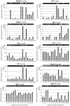
The mRNA expression profiles of differentially expressed up-regulated genes. Gel images on top show PCR product bands stained with ethidium bromide; band intensities are also presented graphically below for clarity. Lane numbers 1 to 8 and 9 to 16 indicate the 6 and 24 h samples (1/9 = sIC, saline Ischemic Core; 2/10 = pacapIC, PACAP38 Ischemic Core; 3/11 = sIP, saline Ischemic Penumbra; 4/12 = pacapIP, PACAP38 Ischemic Penumbra; 5/13 = sHC, saline Healthy Core; 6/14 = pacapHC, PACAP38 Healthy Core; 7/15 = sHP, saline Healthy Penumbra; 8/16 = pacapHP, PACAP38 Healthy Penumbra), respectively. Semi-quantitative RT-PCR was performed as described in Materials and Methods, and the gene-specific primers are detailed in Table 1.
Figure 4
Figure 4
The mRNA expression profiles of differentially expressed down-regulated genes. Gel images on top show PCR product bands stained with ethidium bromide; band intensities are also presented graphically below for clarity. Lane numbers 1 to 8 and 9 to 16 indicate the 6 and 24 h samples (1/9 = sIC, saline Ischemic Core; 2/10 = pacapIC, PACAP38 Ischemic Core; 3/11 = sIP, saline Ischemic Penumbra; 4/12 = pacapIP, PACAP38 Ischemic Penumbra; 5/13 = sHC, saline Healthy Core; 6/14 = pacapHC, PACAP38 Healthy Core; 7/15 = sHP, saline Healthy Penumbra; 8/16 = pacapHP, PACAP38 Healthy Penumbra), respectively. Semi-quantitative RT-PCR was performed as described in Materials and Methods, and the gene-specific primers are detailed in Table 1.
Figure 5
Figure 5
Western blot analysis of the CRMP2 protein cross-reacting proteins in ischemic core and penumbra of ipsilateral (right) hemisphere along with their abundance in corresponding regions in the healthy contralateral hemisphere (left). In (A), lane numbers 1 to 8 and in (B), lane numbers 9 to 16 indicate the 6 and 24 h samples, respectively. Abbreviations are the same as in Figure 2 legend. Proteins cross-reacting with the anti-CRMP2 protein antibody are visible as three constitutively present proteins (~70, ~65, and ~63 kDa size) in all samples. The ~56 kDa cross-reacting protein is seen not only in the ischemic core but also in the penumbra. Total protein extraction, separation, Western blotting, and image analyses procedures are detailed in Materials and Methods.

Similar articles

Cited by

References

    1. Gusev E.I., Skvortsova V.I. Brain Ischemia. Kluwer Academic/Plenum Publishers; New York, NY, USA: 2003.
    1. Kimura C., Ohkubo S., Ogi K., Hosoya M., Itoh Y., Onda H., Miyata A., Jian L., Dahl R.R., Stibbs H.H., et al. A novel peptide which stimulates adenylate cyclase: Molecular cloning and characterization of the ovine and human cDNAs. Biochem. Biophys. Res. Commun. 1990;166:81–89. - PubMed
    1. Arimura A. Perspectives on pituitary adenylate cyclase activating polypeptide (PACAP) in the neuroendocrine, endocrine, and nervous systems. Jpn. J. Physiol. 1998;48:301–331. - PubMed
    1. Shioda S., Ozawa H., Dohi K., Mizushima H., Matsumoto K., Nakajo S., Takaki A., Zhou C.J., Nakai Y., Arimura A. PACAP protects hippocampal neurons against apoptosis: Involvement of JNK/SAPK signaling pathway. Ann. N. Y. Acad. Sci. 1998;865:111–117. - PubMed
    1. Ohtaki H., Funahashi H., Dohi K., Oguro T., Horai R., Asano M., Iwakura Y., Yin L., Matsunaga M., Goto N., et al. Suppression of oxidative neuronal damage after transient middle cerebral artery occlusion in mice lacking interleukin-1. Neurosci. Res. 2003;45:313–324. - PubMed

Publication types

MeSH terms

Substances