. 2014 Sep 26;345(6204):1616-20.
doi: 10.1126/science.1255514.

A critical time window for dopamine actions on the structural plasticity of dendritic spines

Affiliations

A critical time window for dopamine actions on the structural plasticity of dendritic spines

Sho Yagishita et al. Science. .

Abstract

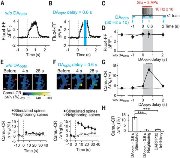
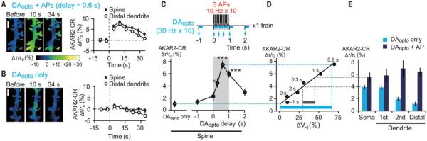
Animal behaviors are reinforced by subsequent rewards following within a narrow time window. Such reward signals are primarily coded by dopamine, which modulates the synaptic connections of medium spiny neurons in the striatum. The mechanisms of the narrow timing detection, however, remain unknown. Here, we optically stimulated dopaminergic and glutamatergic inputs separately and found that dopamine promoted spine enlargement only during a narrow time window (0.3 to 2 seconds) after the glutamatergic inputs. The temporal contingency was detected by rapid regulation of adenosine 3',5'-cyclic monophosphate in thin distal dendrites, in which protein-kinase A was activated only within the time window because of a high phosphodiesterase activity. Thus, we describe a molecular basis of reinforcement plasticity at the level of single dendritic spines.


Figures

Fig. 1
Fig. 1. A temporal profile of dopamine actions on spine enlargement
(A) Injection of AAV vectors for ChR2 and the D1R-MSN marker (PPTA-mCherry) in 3-week-old DAT-Cre mice. (B) Selective stimulation of dopaminergic and glutamatergic inputs by means of blue laser field irradiation to ChR2 and two-photon uncaging of caged-glutamate at a single spine, respectively, in acute slices of NAc obtained from 5- to 7- week-old mice. (C) An amperometric measurement of dopamine (top) by carbon-fiber electrode and whole-cell recording of glutamate-induced current (bottom, 2pEPSP) in identified D1R-MSNs. (D) An STDP protocol with dopamine puff application. (E) Images of the dendritic spine (red arrowhead) that received STDP stimulation in the presence of dopamine (100 μM). (F and G) Time courses of spine enlargement in the presence [(F), 13 spines, 4 dendrites] and absence of dopamine [(G), 58 spines, 14 dendrites]. (H) Amplitudes of spine enlargements with or without dopamine. **P = 0.0041 by Mann-Whitney U test. (I) STDP with repetitive activation of dopaminergic fibers containing ChR2 (blue lines) at 30 Hz, 10 times (DAopto). (J) Images of the dendritic spine (arrowhead) that received STDP + DAopto with a delay of 1 s. (K to M) Time courses of spine enlargement induced by STDP + DAopto at 1 s [(K), 48 spines, 14 dendrites], −1 s [(L), 20 spines, 5 dendrites] and 5 s [(M), 28 spines, 7 dendrites] after STDP onset. (N) Timings of DAopto application. (O) Increases in spine volumes by STDP + DAopto plotted versus DAopto delay (fig. S2, A to C). Data are presented as mean ± SEM. P = 4.2 × 10⁻⁶ with Kruskal-Wallis and **P = 0.0018 (0.6 s) and 0.0027 (1 s) by Steel test in comparison with STDP in the absence of DAopto. Scale bars, 1 μm.
Fig. 2
Fig. 2. Pharmacology of spine enlargement induced by STDP plus DAopto with a 0.6-s delay
(A) Time courses of spine enlargement induced by STDP + DAopto with a 0.6-s delay in the absence (control, 24 spines, 7 dendrites) and presence of NMDAR antagonist (50 μM D-AP5, 22 spines, 6 dendrites), CaMKII inhibitor (3 μM KN62, 23 spines, 6 dendrites), or protein synthesis inhibitor (5 μM anisomycin, 25 spines, 6 dendrites). (B) Time courses of spine enlargement in the presence of D1R antagonist (3 μM SCH23390, 23 spines, 6 dendrites), D2R antagonist (10 μM sulpiride, 22 spines, 6 dendrites), or PKA inhibitor (10 μM PKI, in the pipette, 24 spines, 6 dendrites). (C) Time courses of spine enlargement in the presence of inhibitory (100 μM, in the pipette, 24 spines, 6 dendrites) or control peptide for DARPP-32 (100 μM, in the pipette, 24 spines, 6 dendrites). (D) Averaged volume changes in the absence and presence of the compounds. Data are presented as mean ± SEM. P = 3.4 × 10⁻⁶ with Kruskal-Wallis and *P = 0.023 (AP5), 0.023 (KN62), 0.037 (AIP) (fig. S5A), 0.023 (anisomycin), 0.035 (SCH23390), 0.023 (PKI), 0.037 (KT5720) (fig. S5A), and 0.023 (DARPP-32 inhibitory peptide) with Steel test.
Fig. 3
Fig. 3. DAopto effects on STDP stimulation-induced increases in [Ca2+]i and CaMKII activities
(A and B) Increases in Fluo4-FF fluorescence, representing [Ca2+]i increases, within single spines in response to a train of STDP stimulation in the absence [(A), 20 spines, 15 dendrites] and presence of DAopto with a 0.6-s delay [(B), 15 spines, 8 dendrites]. Blue laser irradiation during DAopto is blanked by the blue bar. (C) STDP and DAopto protocols for Ca2+ and CaMKII imaging. Unlike plasticity induction (Fig. 1N), only one train was applied. (D) No effect of DAopto on the peak values of [Ca2+]i (fig. S6, A to C). P = 0.91 with Kruskal-Wallis test. (E and F) Ratiometric imaging with Camuiα-CR during STDP stimulation in the absence [(E), 33 spines, 14 dendrites] or presence of DAopto with a 0.6-s delay [(F), 42 spines, 14 dendrites]. Relative increases in the ratio are shown as pseudocolor coding in (E). (Bottom) Time courses of FRET ratios in the spines stimulated with glutamate uncaging or the neighboring spines. Scale bars, 1 μm. (G) Dependence of the peak Camuiα-CR FRET ratios on the DAopto delay (fig. S6, D to F). P = 1.3 × 10⁻⁵ with Kruskal-Wallis test and ***P = 8.4 × 10⁻⁵ with Steel test versus those without DAopto. (H) Normalized increases in Camuiα-CR ratios by STDP + DAopto with a 0.6-s delay in the stimulated (42 spines, 14 dendrites) and neighboring spines (42 spines, 14 dendrites), and in the presence of DARPP-32 inhibitory peptide in the stimulated spines (43 spines, 10 dendrites) (fig. S6G). Data are presented as mean ± SEM. P = 8.8 × 10⁻⁹ with Kruskal-Wallis test and *** P = 2.6 × 10⁻⁹ and 1.1 × 10⁻⁶ with Steel test.
Fig. 4
Fig. 4. AP effects on DAopto-induced PKA activation in proximal and distal dendrites
(A and B) Images and time courses of FRET ratios of AKAR2-CR in the spine and distal dendrites stimulated by APs + DAopto with a 0.6-s delay [(A), 153 spines, 25 dendrites] or DAopto only [(B), 158 spines, 26 dendrites]. The relative increases in the ratio were pseudocolor coded as shown in (A). Scale bar, 2 μm. (C) AKAR2-CR responses to APs + DAopto with various delays (fig. S7, E to I). P = 5.3 × 10⁻¹⁰ with Kruskal-Wallis test and *P = 0.012, ***P = 1.3 × 10⁻⁶ (0.6 s), and 4.0 × 10⁻⁴ (1 s) with Steel test versus DAopto only. (D) AKAR2-CR response (C) plotted against spine volume changes (Fig. 1O) for various DAopto timings. The Spearman’s correlation coefficient was 0.94, and P = 0.0048. The blue and gray bars indicate the dynamic ranges of volume changes predicted by the dynamic ranges of AKAR2 responses at dendritic spine (blue) and soma (gray). (E) AKAR2-CR responses at the soma and first, second, and distal dendrites in response to DAopto only and DAopto + APs with a 0.6-s delay (fig. S8, A and B).

Comment in

References

    1. Thorndike EL. Animal Intelligence. Macmillan; New York: 1911.
    1. Pavlov IP. Conditioned Reflexes; An Investigation of the Physiological Activity of the Cerebral Cortex. Oxford Univ. Press; Humphrey Milford, London: 1927. - PMC - PubMed
    1. Roitman MF, Wheeler RA, Carelli RM. Nucleus accumbens neurons are innately tuned for rewarding and aversive taste stimuli, encode their predictors, and are linked to motor output. Neuron. 2005;45:587–597. - PubMed
    1. Dan Y, Poo MM. Spike timing-dependent plasticity: From synapse to perception. Physiol. Rev. 2006;86:1033–1048. - PubMed
    1. Black J, Belluzzi JD, Stein L. Reinforcement delay of one second severely impairs acquisition of brain self-stimulation. Brain Res. 1985;359:113–119. - PubMed

Publication types

MeSH terms