. 2014 Sep 26;345(6204):1620-3.
doi: 10.1126/science.1256679.

Ligand binding to the FeMo-cofactor: structures of CO-bound and reactivated nitrogenase

Affiliations

Ligand binding to the FeMo-cofactor: structures of CO-bound and reactivated nitrogenase

Thomas Spatzal et al. Science. .

Abstract

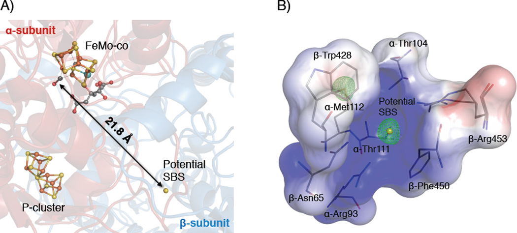
The mechanism of nitrogenase remains enigmatic, with a major unresolved issue concerning how inhibitors and substrates bind to the active site. We report a crystal structure of carbon monoxide (CO)-inhibited nitrogenase molybdenum-iron (MoFe)-protein at 1.50 angstrom resolution, which reveals a CO molecule bridging Fe2 and Fe6 of the FeMo-cofactor. The μ2 binding geometry is achieved by replacing a belt-sulfur atom (S2B) and highlights the generation of a reactive iron species uncovered by the displacement of sulfur. The CO inhibition is fully reversible as established by regain of enzyme activity and reappearance of S2B in the 1.43 angstrom resolution structure of the reactivated enzyme. The substantial and reversible reorganization of the FeMo-cofactor accompanying CO binding was unanticipated and provides insights into a catalytically competent state of nitrogenase.


Figures

Figure 1
Figure 1. CO-inhibited MoFe-protein (Av1-CO)
Refined structure of the CO-bound FeMo-cofactor at a resolution of 1.50 Å. A) View along the Fe1-C-Mo direction. The electron density (2Fo-Fc) map is contoured at 4.0 σ and represented as blue mesh. The density at the former S2B site is significantly decreased and in excellent agreement with bound CO (see also C)). B) Same orientation as A) superimposed with the anomalous density map calculated at 7100 eV (green) at a resolution of 2.1 Å contoured at 4.0 σ showing the significant reduction of anomalous electron density at the CO site. C) Side view of FeMo-cofactor highlighting the μ2 binding geometry of CO. The electron density (2Fo-Fc) map (blue mesh) surrounding CO-Fe2-Fe6-C is contoured at 1.5 σ. D) Same orientation as C) highlighting the ligand environment of the metal center. The catalytically important side chain residues α-Val70 and α-His195 are in close proximity to the CO-binding site. Iron atoms are shown in orange, sulfur in yellow, molybdenum in turquoise, carbon in grey, nitrogen in blue and oxygen in red.
Figure 2
Figure 2. Reactivated MoFe-protein (Av1-reactivated)
Refined structure of the FeMo-cofactor at a resolution of 1.43 Å. A) View along the Fe1-C-Mo direction. The electron density (2Fo-Fc) map is contoured at 4.0 σ and represented as blue mesh. Electron density at the S2B site is in excellent agreement with a regained sulfur. B) Same orientation as A) superimposed with the anomalous density map (green) at a resolution of 2.15 Å contoured at 4.0 σ showing the presence of anomalous density at the S2B site. Color scheme is according to Figure 1.
Figure 3
Figure 3. Overview of the potential sulfur binding site (SBS) in the CO-inhibited MoFe-protein (Av1-CO)
A) Location of the potentially bound sulfur in a protein cavity on the interface between the α- and β-subunit of the α2β2 MoFe-protein. The potential SBS is located 22 Å away from its former position in the FeMo-cofactor (S2B-site). B) Close-up view on the binding cavity. Positive surface charge is represented in blue, negative surface charge in red. The anomalous density map at a resolution of 2.1 Å is represented as green mesh and contoured at 4.0 σ showing the presence of anomalous density at the potential SBS. The side chain sulfur of α-Met112 provides an internal standard for full occupancy. The color scheme is according to Figure 1.

Comment in

References

    1. Burgess BK, Lowe DJ. Chem. Rev. 1996;96:2983–3011. - PubMed
    1. Howard JB, Rees DC. Proc. Natl. Acad. Sci. USA. 2006;103:17088–17093. - PMC - PubMed
    1. Seefeldt LC, Hoffman BM, Dean DR. Ann. Rev. Biochem. 2009;78:701–722. - PMC - PubMed
    1. Georgiadis MM, Komiya H, Chakrabarti P, Woo D, Kornuc JJ, Rees DC. Science. 1992;257:1653–1659. - PubMed
    1. Kim JS, Rees DC. Nature. 1992;360:553–560. - PubMed

Publication types