. 2014 Nov 21;289(47):32559-70.
doi: 10.1074/jbc.M114.602599. Epub 2014 Sep 25.

A peptide derived from G0/G1 switch gene 2 acts as noncompetitive inhibitor of adipose triglyceride lipase

Affiliations

A peptide derived from G0/G1 switch gene 2 acts as noncompetitive inhibitor of adipose triglyceride lipase

Ines K Cerk et al. J Biol Chem. .

Abstract

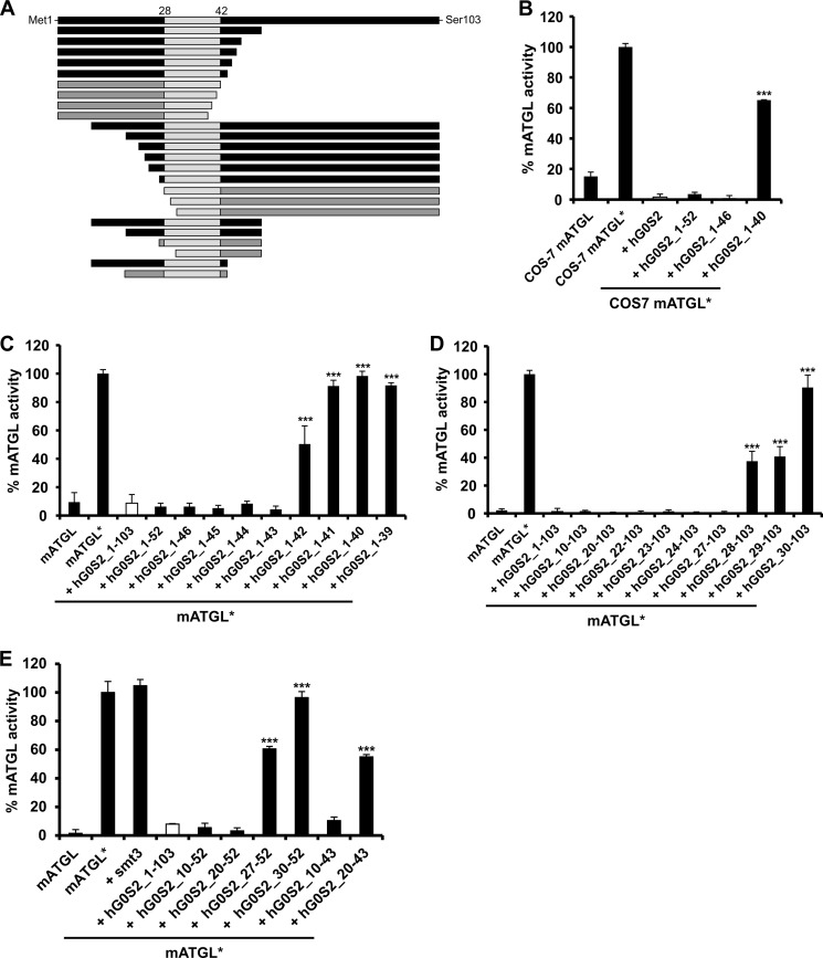
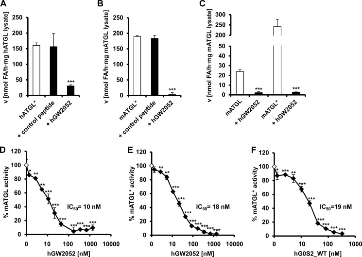
The protein G0/G1 switch gene 2 (G0S2) is a small basic protein that functions as an endogenous inhibitor of adipose triglyceride lipase (ATGL), a key enzyme in intracellular lipolysis. In this study, we identified a short sequence covering residues Lys-20 to Ala-52 in G0S2 that is still fully capable of inhibiting mouse and human ATGL. We found that a synthetic peptide corresponding to this region inhibits ATGL in a noncompetitive manner in the nanomolar range. This peptide is highly selective for ATGL and does not inhibit other lipases, including hormone-sensitive lipase, monoacylglycerol lipase, lipoprotein lipase, and patatin domain-containing phospholipases 6 and 7. Because increased lipolysis is linked to the development of metabolic disorders, the inhibition of ATGL by G0S2-derived peptides may represent a novel therapeutic tool to modulate lipolysis.

Keywords: Adipose Triglyceride Lipase (ATGL); Atglistatin; Enzyme Inhibitor; G0/G1 Switch Gene 2 (G0S2); Lipid; Lipid Metabolism; Lipolysis; Noncompetitive Inhibition; Peptides; hGW2052.


Figures

FIGURE 1.
FIGURE 1.
Minimal sequence requirements of human G0S2 for ATGL inhibition. A, graphical representation of full-length and shortened versions of hG0S2, those capable of inhibiting ATGL activity are illustrated in black. The largely hydrophobic sequence stretch from Val-28 to Met-42 is depicted in light gray. B–E, activity assays were performed in the presence of CGI-58 as indicated by an asterisk (ATGL*). G0S2 and variants thereof were expressed in E. coli and added as cell lysates. Overexpressed mATGL from E. coli lysates was used for all assays with the exception of the assay shown in B. B, inhibition of TG hydrolase activity of mATGL containing COS-7 cell extracts by WT and C-terminally truncated hG0S2 variants. C, inhibition of TG hydrolase activity of mATGL by WT and C-terminally truncated hG0S2 variants. D, inhibition of TG hydrolase activity of mATGL by WT hG0S2 and variants with N-terminal deletions. E, effect of different N- and C-terminal truncations in hG0S2 on mATGL activity. The fusion tag smt3 is shown as control.
FIGURE 2.
FIGURE 2.
Inhibition of ATGL by the peptide hGW2052 corresponding to residues Lys-20 to Ala-52 of hG0S2. Activity assays performed in the presence of CGI-58 are indicated by an asterisk (ATGL*). A and B, TG hydrolase activity assays of CGI-58 activated ATGL in the absence and in the presence of 1.4 μm hGW2052. A peptide harboring a sequence unrelated to G0S2 served as negative control. A, TG hydrolase activity of hATGL* expressed in COS-7 cells. B, TG hydrolase activity of mATGL* expressed in E. coli. C–F, mATGL used for these assays was overexpressed in E. coli. C, inhibition of the TG hydrolase activity of mATGL- and CGI-58-stimulated mATGL by 1.4 μm of the peptide hGW2052. D, dose-dependent inhibition of TG hydrolase activity of mATGL. E, mCGI-58-stimulated mATGL by the peptide hGW2052. F, dose-dependent inhibition of CGI-58-stimulated mATGL by WT hG0S2. Statistical significance was assigned according to the following scheme: *, p < 0.05; **, p < 0.01; ***, p < 0.001.
FIGURE 3.
FIGURE 3.
hGW2052 inhibits ATGL via a noncompetitive mechanism. All TG hydrolase activity assays in this figure were performed with mATGL expressed in E. coli and in the presence of CGI-58 (ATGL*). A, time dependence of FA release during TG hydrolase activity assay. B, reaction velocity of mATGL lysates depends on the concentration of triolein substrate. C, relationship between TG hydrolase reaction rates and substrate concentration (within the linear concentration range) at varying hGW2052 concentrations. D, inhibition of mATGL activity in the presence of different hGW2052 concentrations with increasing concentrations of triolein substrate. E, Dixon-Plot for kinetic analysis of mATGL inhibition by hGW2052. The reciprocal velocity is plotted against the concentration of hGW2052 at various substrate concentrations. In noncompetitive inhibition, the lines converge on the x axis with the intersection point giving −Ki. The inset shows nonlinear regression analysis (GraphPad Prism 5) using the equation for noncompetitive inhibition.
FIGURE 4.
FIGURE 4.
The Peptide hGW2052 selectively inhibits ATGL. TG hydrolase activity of ATGL, HSL, and lipoprotein lipase (LPL) was measured using triolein as substrate. MGL activity was determined using rac-(1,3)-monooleylglycerol as substrate. PNPLA6 and PNPLA7 activities were detected in the presence of 1-oleoyl-2-hydroxy-sn-glycero-3-phosphocholine. A, inhibition of TG hydrolase activity in WAT lysates of overnight fasted WT and ATGL-KO mice by 1.4 μm hGW2052, 40 μm Atglistatin, and 25 μm of the HSL inhibitor Hi 76-0079, respectively. Dose-dependent effect of hGW2052 and control peptide is shown on murine PNPLA6 (B), PNPLA7 lysophospholipase activities (C), murine MGL monoacylglycerol hydrolase activity (D), HSL triglyceride hydrolase activity (E), and bovine lipoprotein lipase triglyceride hydrolase activity (F).

References

    1. Li L. O., Klett E. L., Coleman R. A. (2010) Acyl-CoA synthesis, lipid metabolism and lipotoxicity. Biochim. Biophys. Acta 1801, 246–251 - PMC - PubMed
    1. Samuel V. T., Petersen K. F., Shulman G. I. (2010) Lipid-induced insulin resistance: unravelling the mechanism. Lancet 375, 2267–2277 - PMC - PubMed
    1. Unger R. H., Clark G. O., Scherer P. E., Orci L. (2010) Lipid homeostasis, lipotoxicity and the metabolic syndrome. Biochim. Biophys. Acta 1801, 209–214 - PubMed
    1. Unger R. H., Scherer P. E. (2010) Gluttony, sloth and the metabolic syndrome: a roadmap to lipotoxicity. Trends Endocrinol. Metab. 21, 345–352 - PMC - PubMed
    1. Karpe F., Dickmann J. R., Frayn K. N. (2011) Fatty acids, obesity, and insulin resistance: time for a reevaluation. Diabetes 60, 2441–2449 - PMC - PubMed

Publication types

MeSH terms

LinkOut - more resources