. 2014 Nov 7;289(45):31212-23.
doi: 10.1074/jbc.M114.598177. Epub 2014 Sep 25.

Torque generation of Enterococcus hirae V-ATPase

Affiliations

Torque generation of Enterococcus hirae V-ATPase

Hiroshi Ueno et al. J Biol Chem. .

Abstract

V-ATPase (V(o)V1) converts the chemical free energy of ATP into an ion-motive force across the cell membrane via mechanical rotation. This energy conversion requires proper interactions between the rotor and stator in V(o)V1 for tight coupling among chemical reaction, torque generation, and ion transport. We developed an Escherichia coli expression system for Enterococcus hirae V(o)V1 (EhV(o)V1) and established a single-molecule rotation assay to measure the torque generated. Recombinant and native EhV(o)V1 exhibited almost identical dependence of ATP hydrolysis activity on sodium ion and ATP concentrations, indicating their functional equivalence. In a single-molecule rotation assay with a low load probe at high ATP concentration, EhV(o)V1 only showed the "clear" state without apparent backward steps, whereas EhV1 showed two states, "clear" and "unclear." Furthermore, EhV(o)V1 showed slower rotation than EhV1 without the three distinct pauses separated by 120° that were observed in EhV1. When using a large probe, EhV(o)V1 showed faster rotation than EhV1, and the torque of EhV(o)V1 estimated from the continuous rotation was nearly double that of EhV1. On the other hand, stepping torque of EhV1 in the clear state was comparable with that of EhV(o)V1. These results indicate that rotor-stator interactions of the V(o) moiety and/or sodium ion transport limit the rotation driven by the V1 moiety, and the rotor-stator interactions in EhV(o)V1 are stabilized by two peripheral stalks to generate a larger torque than that of isolated EhV1. However, the torque value was substantially lower than that of other rotary ATPases, implying the low energy conversion efficiency of EhV(o)V1.

Keywords: Bioenergetics; Membrane Protein; Molecular Motor; Single-molecule Biophysics; V-ATPase; Vacuolar ATPase.


Figures

FIGURE 1.
FIGURE 1.
Gel electrophoresis. Lanes 1–4, SDS-PAGE of recombinant EhV1, reconstituted EhV1, native EhVoV1, and recombinant EhVoV1. A 16% gel was used, and 2 pmol of protein was loaded in each lane. The molecular masses of the a, A, B, d, D, E, c, G, and F subunits are 76, 66, 51, 38, 25, 23, 16, 14, and 11 kDa, respectively. The F subunit of reconstituted EhV1 has an additional linker sequence for purification of the DF subcomplex and caused a band shift to a higher molecular mass. Lanes 5–8, immunoblots stained by alkaline phosphatase-streptavidin conjugates, showing biotin labeling of the D subunit of reconstituted EhV1 and the A subunit of recombinant EhVoV1. Lane 5, nonbiotinylated recombinant EhV1. Lane 6, reconstituted EhV1 containing the biotinylated D subunit. Lane 7, nonbiotinylated native EhVoV1. Lane 8, recombinant EhVoV1 containing the biotinylated A subunit. Lanes 9 and 10, SDS-PAGE of native and recombinant EhVoV1, respectively. A 9% gel was used to resolve the band of the a subunit; 1 pmol protein was loaded in each lane. The a subunit migrated slightly faster than the A subunit as reported previously (15).
FIGURE 2.
FIGURE 2.
Biochemical assay of the ATPase activity of EhVoV1. A, dependence of ATPase activity on Na+ concentration at 5 mm ATP. Average ATPase rates of native (open circles) and recombinant (open triangles) EhVoV1 are shown (n ≥ 3). The error bars represent standard deviations. The inset shows the expanded plot for lower Na+ concentrations. Solid and dashed curves show the fit of the model with a sum of two Michaelis-Menten equations assuming two independent binding sites of Na+: V = Vmax1[Na+]/(Km1Na + [Na+]) + Vmax2[Na+]/(Km2Na + [Na+]). The Km1Na, Km2Na, Vmax1, and Vmax2 values were 20 ± 5 μm, 42 ± 8 mm, 38 ± 1 s−1, and 55 ± 2 s−1 for native EhVoV1 and 13 ± 3 μm, 50 ± 9 mm, 37 ± 1 s−1, and 49 ± 2 s−1 for recombinant EhVoV1 (fitted value ± S.E. of the fit), respectively. B, double-reciprocal plots of A. C, dependence of ATPase activity on ATP concentration at high Na+ concentration (300 mm). Averages of one-third of the ATPase rate (corresponding to the rotation rate) for native (open circles) and recombinant (open triangles) EhVoV1 determined by a biochemical assay at pH 8.5 (n ≥ 3) are shown. The solid and dashed lines indicate the fit with the Michaelis-Menten equation: V = VmaxATP × [ATP]/(KmATP + [ATP]). The values of KmATP, VmaxATP, and the second order binding rate constant for ATP (3 × VmaxATP/KmATP) are shown in Table 2. D, inhibitory effect of DCCD on ATPase activity of detergent-solubilized recombinant EhVoV1. After incubation of recombinant EhVoV1 with 200 μm DCCD for indicated times, residual ATPase activity was examined (n ≥ 3) under the same buffer condition as the rotation assay at 4 mm ATP without (open triangles) or with 0.15% LDAO (filled triangles). Residual ATPase activities decreased to 24 ± 3 and 21 ± 3% after 60 and 90 min of incubation with DCCD, respectively (mean ± S.D.). Residual ATPase activities were recovered to 90 ± 1 and 88 ± 3% by the addition of LDAO, even after 60 and 90 min of incubation with DCCD, respectively. The error bars represent standard deviations.
FIGURE 3.
FIGURE 3.
Rotation rate of EhVoV1 and EhV1 at saturating ATP concentration and ATP concentration dependence of the ATPase activity. A, schematic image of the rotation assay of EhV1 (left panel) and EhVoV1 (right panel). The A3B3 ring of EhV1 or the c ring of EhVoV1 was fixed on the Ni2+-NTA glass surface with a His tag. A streptavidin-coated 40-nm gold colloid was attached to the biotinylated D subunit in EhV1 or the A subunit in EhVoV1. B, rotation rate at saturating ATP concentration and ATP concentration dependence of the ATPase activity. Average rotation rates for recombinant EhVoV1 (open red triangle, 45 ± 12 rps, mean ± S.D., n = 15) and reconstituted EhV1 (open blue triangle, 102 ± 13 rps, n = 6) determined by a rotation assay using a low load probe at 4 mm ATP and pH 6.5 are shown. Filled red and blue triangles indicate rotation rates for individual molecules. Averages of one-third of the ATPase activity (corresponding to the rotation rate) for recombinant EhVoV1 (open red circles) and reconstituted EhV1 (open blue circles) determined by a biochemical assay at pH 6.5 (n ≥ 3) are also shown. The solid lines indicate the fit with the Michaelis-Menten equation: V = VmaxATP× [ATP]/(KmATP + [ATP]). The values of KmATP, VmaxATP, and the second order binding rate constant for ATP (3 × VmaxATP/KmATP) are shown in Table 2.
FIGURE 4.
FIGURE 4.
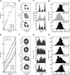
Low load rotation of EhVoV1 and EhV1. A and E, typical time course of rotation of EhV1 at 3 mm ATP recorded at 10,000 fps (29) and of EhVoV1 at 4 mm ATP in the presence of 300 mm NaCl recorded at 5,000–10,000 fps by using a 40-nm gold colloid. The time course of EhV1 includes two reversible distinct states: clear (black) and unclear (gray) (29). B and F, the x-y trajectories of the centroid of the rotating gold colloid shown in A and E. C and G, the distribution of the rotary angle shown in B and F. D and H, the distribution of the distance of the centroid of the gold colloid from the rotation center. Average distance in unclear state (gray, ∼16 nm) is distinctly shorter than those in the clear state (black, 18–20 nm), and the histogram in the unclear state is distributed closer to the rotation center (∼13% within 10 nm) than that in clear state (<5% within 10 nm). The numbers in A–H indicate corresponding parts and molecules.
FIGURE 5.
FIGURE 5.
Rotation rate and torque of EhVoV1 and EhV1 determined by continuous rotation of a large probe. Rotation rate (A) and torque (B) estimated by FT analysis of EhVoV1 (columns 1 and 2), EhV1 (columns 3–5), and TF1 (columns 6). The rotation of EhVoV1 was observed at 50 mm KCl (column 1, n = 39) or 300 mm NaCl (columns 2, n = 25) in the presence of 0.05% DDM with 287-nm duplex beads. The rotation of EhV1 was observed at 50 mm KCl in the absence (columns 3, n = 9) or presence (columns 4, n = 4) of 0.05% DDM with 287-nm duplex beads or with 200-nm duplex beads (columns 5, n = 11). The rotation of TF1 was observed at 50 mm KCl without DDM with 287-nm duplex beads (columns 6, n = 8). ATP concentration was 4 mm in all conditions. Filled circles indicate rotation rates or torques for individual molecules. Columns show the averages. The error bars indicate standard deviations.
FIGURE 6.
FIGURE 6.
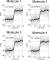
Stepping torque of EhV1 in clear rotation state. Time courses of the stepping rotation of four single EhV1 molecules at 3 mm ATP, captured at a rate of 300,000–372,000 fps. Each gray line shows 18 successive steps superimposed at the point closest to the midpoint of each 120° step, whereas black lines and dots show the averages of the 18 lines. The dotted lines show the fit with the linear function.

Similar articles

Cited by

References

    1. Nishi T., Forgac M. (2002) The vacuolar H+-ATPases: nature's most versatile proton pumps. Nat. Rev. Mol. Cell Biol. 3, 94–103 - PubMed
    1. Forgac M. (2007) Vacuolar ATPases: rotary proton pumps in physiology and pathophysiology. Nat. Rev. Mol. Cell Biol. 8, 917–929 - PubMed
    1. Bowman E. J., Bowman B. J. (2005) V-ATPases as drug targets. J. Bioenerg. Biomembr. 37, 431–435 - PubMed
    1. Marshansky V., Futai M. (2008) The V-type H+-ATPase in vesicular trafficking: targeting, regulation and function. Curr. Opin. Cell Biol. 20, 415–426 - PMC - PubMed
    1. Stewart A. G., Laming E. M., Sobti M., Stock D. (2014) Rotary ATPases: dynamic molecular machines. Curr. Opin. Struct. Biol. 25C, 40–48 - PubMed

Publication types

LinkOut - more resources