. 2014 Sep 18:7:401-11.
doi: 10.2147/JMDH.S68308. eCollection 2014.

Clinical and symptomatological reflections: the fascial system

Affiliations

Clinical and symptomatological reflections: the fascial system

Bruno Bordoni et al. J Multidiscip Healthc. .

Abstract

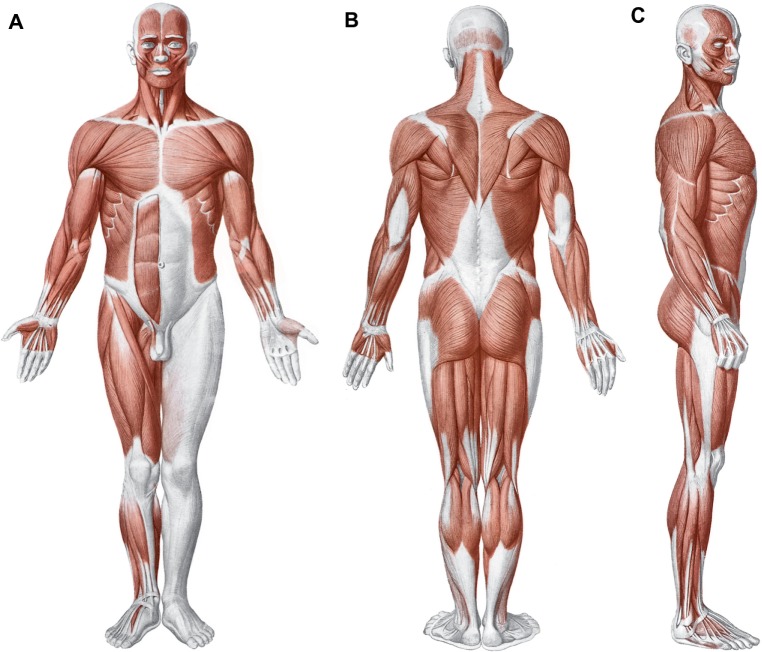
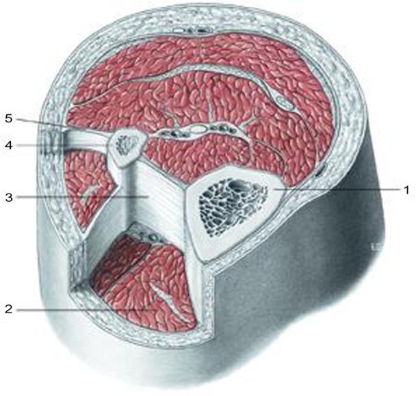
Every body structure is wrapped in connective tissue, or fascia, creating a structural continuity that gives form and function to every tissue and organ. Currently, there is still little information on the functions and interactions between the fascial continuum and the body system; unfortunately, in medical literature there are few texts explaining how fascial stasis or altered movement of the various connective layers can generate a clinical problem. Certainly, the fascia plays a significant role in conveying mechanical tension, in order to control an inflammatory environment. The fascial continuum is essential for transmitting muscle force, for correct motor coordination, and for preserving the organs in their site; the fascia is a vital instrument that enables the individual to communicate and live independently. This article considers what the literature offers on symptoms related to the fascial system, trying to connect the existing information on the continuity of the connective tissue and symptoms that are not always clearly defined. In our opinion, knowing and understanding this complex system of fascial layers is essential for the clinician and other health practitioners in finding the best treatment strategy for the patient.

Keywords: fascia; low back pain; neck; osteopathic; pain.


Figures

Figure 1
Figure 1
Shape and arrangement of the muscles on the ventral (A), dorsal (B), and lateral (C) surface of the human body. Notes: Copyright Edi.Ermes, Milano. Reproduced with permission from Anastasi et al. AA VV, Anatomia dell’uomo, 4th ed, Edi.Ermes, Milano [Human anatomy].
Figure 2
Figure 2
Transverse section at the level of the upper third of the leg. Notes: 1, tibia; 2, muscular loggia; 3, interosseous membrane; 4, fibula; 5, intermuscular septum. All tissues are enveloped by fascial continuum. Copyright Edi.Ermes, Milano. Reproduced with permission from Anastasi et al. AA VV, Anatomia dell’uomo, 4th ed, Edi.Ermes, Milano [Human anatomy].
Figure 3
Figure 3
Presentation of the mimic muscles of the head. These muscles occupy a superficial position. Notes: 1, frontal muscle; 2, orbicularis oculi muscle, part of the eyelid; 3, procero muscle; 4, medial palpebral ligament; 5, nasal muscle; 6, elevator muscle of the upper lip and of the wing of nose muscle; 7, elevator of the upper lip muscle; 8, small zygomatic muscle; 9, great zygomatic muscle; 10, orbicularis oris muscle; 11, risorio muscle; 12, squared muscle of the inferior lip; 13, mentalis muscle; 14, triangular muscle; 15, platysma muscle; 16, parotid fascia; 17, fascia masseter; 18, anterior auricular muscle; 19, occipital muscle; 20, temporoparietal and upper auricular muscles; 21, aponeurotic galea. Copyright Edi.Ermes, Milano. Reproduced with permission from Anastasi et al. AA VV, Anatomia dell’uomo, 4th ed, Edi.Ermes, Milano [Human anatomy].
Figure 4
Figure 4
Diagram showing the elevator muscle of the upper eyelid and extrinsic muscle of the eyeball, after lifting the cranial vault and the lateral wall of the orbit. Notes: 1, Superior oblique muscle; 2, optic nerve; 3, superior rectus muscle; 4, tendinous ring of Zinn; 5, inferior rectus muscle; 6, inferior oblique muscle; 7, lateral rectus muscle; 8, superioris levator palpebrae muscle. Copyright Edi. Ermes, Milano. Reproduced with permission from Anastasi et al. AA VV, Anatomia dell’uomo, 4th ed, Edi.Ermes, Milano [Human anatomy].
Figure 5
Figure 5
Aspect of the pericardium after resection and removal of its anterior wall, the great vessels, and heart. Notes: In evidence the base of the heart, the posterior wall and the areas of folding of the serous pericardium at the level of the great vessels. 1, left clavicle; 2, left external jugular vein; 3, left subclavian artery; 4, left internal jugular vein; 5, sternal manubrium; 6, left subclavian artery; 7, left common carotid artery; 8, carotid arch; 9, pulmonary trunk; 10, right pulmonary artery; 11, left pulmonary artery; 12, left lung; 13, left upper pulmonary vein; 14, left inferior pulmonary vein; 15, posterior wall; 16, left mediastinal pleura; 17, pericardial sac; 18, diaphragm; 19, base; 20, pericardial sac (dissected); 21, inferior vena cava; 22, right lower pulmonary vein; 23, right mediastinal pleura; 24, right lung; 25, diverticulum Haller; 26, upper right pulmonary vein; 27, transverse sinus of the pericardium; 28, visceral layer; 29, serous pericardium, parietal layer; 30, apex of the pericardial sac; 31, superior vena cava; 32, brachiocephalic trunk; 33, right first rib; 34, left common carotid artery; 35, right common artery; 36, right subclavian vein; 37, right internal jugular vein. Copyright Edi.Ermes, Milano. Reproduced with permission from Anastasi et al. AA VV, Anatomia dell’uomo, 4th ed, Edi.Ermes, Milano [Human anatomy].
Figure 6
Figure 6
Schematic representation of the front section of the nasal cavity viewed from the rear face. Notes: The section captures the outlet of the maxillary sinus into the nasal cavity (red arrow). 1, frontal sinus; 2, cribriform plate; 3, crista galli; 4, ethmoid bone; 5, ethmoidal cells; 6, perpendicular plate; 7, media nasal concha; 8, inferior nasal concha; 9, hard palate; 10, nasal cavity; 11, mucosa of the nasal cavity; 12, mucosa of the maxillary sinus; 13, maxillary sinus; 14, orbital plate; 15, orbit. Copyright Edi. Ermes, Milano. Reproduced with permission from Anastasi et al. AA VV, Anatomia dell’uomo, 4th ed, Edi.Ermes, Milano [Human anatomy].
Figure 7
Figure 7
X-ray shows an implantable cardiac defibrillator/pacemaker following heart surgery.
Figure 8
Figure 8
Pacemaker biventricular-bicameral/implantable cardiac defibrillator. Notes: X-ray shows a slight elevation of the left hemidiaphragm due to a lesion of the phrenic nerve following sternotomy after heart surgery.

References

    1. Bordoni B, Zanier E. Skin, fascias, and scars: symptoms and systemic connections. J Multidiscip Healthc. 2013;7:11–24. - PMC - PubMed
    1. Abbott RD, Koptiuch C, Iatridis JC, Howe AK, Badger GJ, Langevin HM. Stress and matrix-responsive cytoskeletal remodeling in fibroblasts. J Cell Physiol. 2013;228(1):50–57. - PMC - PubMed
    1. Stecco A, Gesi M, Stecco C, Stern R. Fascial components of the myofascial pain syndrome. Curr Pain Headache Rep. 2013;17(8):352. - PubMed
    1. Buckley CD. Why does chronic inflammation persist: An unexpected role for fibroblasts. Immunol Lett. 2011;138(1):12–14. - PMC - PubMed
    1. Bordoni B, Zanier E. Cranial nerves XIII and XIV: nerves in the shadows. J Multidiscip Healthc. 2013;6:87–91. - PMC - PubMed