

The role of microglia in diabetic retinopathy
- PMID: 25258680
- PMCID: PMC4166427
- DOI: 10.1155/2014/705783
The role of microglia in diabetic retinopathy
Abstract
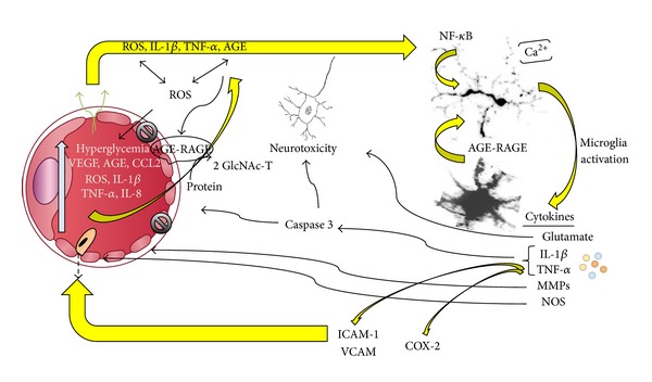
There is growing evidence that chronic inflammation plays a role in both the development and progression of diabetic retinopathy. There is also evidence that molecules produced as a result of hyperglycemia can activate microglia. However the exact contribution of microglia, the resident immune cells of the central nervous system, to retinal tissue damage during diabetes remains unclear. Current data suggest that dysregulated microglial responses are linked to their deleterious effects in several neurological diseases associated with chronic inflammation. As inflammatory cytokines and hyperglycemia disseminate through the diabetic retina, microglia can change to an activated state, increase in number, translocate through the retina, and themselves become the producers of inflammatory and apoptotic molecules or alternatively exert anti-inflammatory effects. In addition, microglial genetic variations may account for some of the individual differences commonly seen in patient's susceptibility to diabetic retinopathy.
Figures
References
-
- Bresnick GH. Diabetic retinopathy viewed as a neurosensory disorder. Archives of ophthalmology. 1986;104(7):989–990. - PubMed
-
- Rungger-Brändle E, Dosso AA, Leuenberger PM. Glial reactivity, an early feature of diabetic retinopathy. Investigative Ophthalmology and Visual Science. 2000;41(7):1971–1980. - PubMed
-
- Lieth E, Gardner TW, Barber AJ, Antonetti DA. Retinal neurodegeneration: early pathology in diabetes. Clinical & Experimental Ophthalmology. 2000;28(1):3–8. - PubMed
Publication types
Grants and funding
LinkOut - more resources
Full Text Sources
Other Literature Sources
Medical
