. 2014 Sep 26;9(9):e108602.
doi: 10.1371/journal.pone.0108602. eCollection 2014.

Melatonin Reverses Fas, E2F-1 and Endoplasmic Reticulum Stress Mediated Apoptosis and Dysregulation of Autophagy Induced by the Herbicide Atrazine in Murine Splenocytes

Affiliations

Melatonin Reverses Fas, E2F-1 and Endoplasmic Reticulum Stress Mediated Apoptosis and Dysregulation of Autophagy Induced by the Herbicide Atrazine in Murine Splenocytes

Shweta Sharma et al. PLoS One. .

Abstract

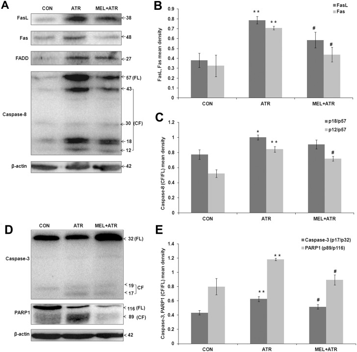
Exposure to the herbicide Atrazine (ATR) can cause immunotoxicity, apart from other adverse consequences for animal and human health. We aimed at elucidating the apoptotic mechanisms involved in immunotoxicity of ATR and their attenuation by Melatonin (MEL). Young Swiss mice were divided into control, ATR and MEL+ATR groups based on daily (x14) intraperitoneal administration of the vehicle (normal saline), ATR (100 mg/kg body weight) and MEL (20 mg/kg body weight) with ATR. Isolated splenocytes were processed for detection of apoptosis by Annexin V-FITC and TUNEL assays, and endoplasmic reticulum (ER) stress by immunostaining. Key proteins involved in apoptosis, ER stress and autophagy were quantified by immunoblotting. ATR treatment resulted in Fas-mediated activation of caspases 8 and 3 and inactivation of PARP1 which were inhibited significantly by co-treatment with MEL. MEL also attenuated the ATR-induced, p53 independent mitochondrial apoptosis through upregulation of E2F-1 and PUMA and suppression of their downstream target Bax. An excessive ER stress triggered by ATR through overexpression of ATF-6α, spliced XBP-1, CREB-2 and GADD153 signals was reversed by MEL. MEL also reversed the ATR-induced impairment of autophagy which was indicated by a decline in BECN-1, along with significant enhancement in LC3B-II and p62 expressions. Induction of mitochondrial apoptosis, ER stress and autophagy dysregulation provide a new insight into the mechanism of ATR immunotoxicity. The cytoprotective role of MEL, on the other hand, was defined by attenuation of ER stress, Fas-mediated and p53 independent mitochondria-mediated apoptosis as well as autophagy signals.


Conflict of interest statement

Competing Interests: The authors have declared that no competing interests exist.

Figures

Figure 1
Figure 1. ATR-induced apoptosis in murine splenocytes was ameliorated by MEL.
(A) Representative images of TUNEL staining in splenocytes of control (CON), atrazine (ATR) and melatonin with atrazine co-treated (MEL+ATR) mice. All cell nuclei were stained with propidium iodide (red fluorescence). TUNEL (fluorescein-12-dUTP labeled fragmented DNA) positive nuclei, displaying green fluorescence, are arrow-marked. Insets show individual and merged fluorescence of single cells at a higher magnification. (B) Histogram showing TUNEL positivity index. Data are expressed as mean ± SEM of 3 experiments (**P<0.01 versus CON; ## P<0.01 versus ATR).
Figure 2
Figure 2. ATR-induced Fas mediated apoptosis was inhibited by MEL.
(A) Representative immunoblots of splenocyte lysates from CON, ATR and MEL+ATR mice. FasL, Fas, FADD, Caspase-8 and β-actin proteins were visualized by chemiluminiscence. Corresponding histograms show (B) FasL, Fas and (C) Caspase-8 (CF/FL; p18/p57 and p12/p57) expressions as mean densities normalized by β-actin density. (D) Representative immunoblots of Caspase-3, PARP1 and β-actin. Corresponding histogram (E) shows caspase-3 (CF/FL, p17/p32) and PARP1 (CF/FL, p89/p116) mean densities normalized by β-actin. Data are presented as mean ± SEM of 3 independent experiments (*P<0.05, **P<0.01 versus CON; # P<0.05 versus ATR). FL, full length; CF, cleaved fragments.
Figure 3
Figure 3. ATR-induced mitochondria mediated apoptosis was inhibited by MEL.
(A) Representative immunoblots of p53, E2F-1, PUMA, Bax, Bcl-2 and β-actin in CON, ATR and MEL+ATR groups. Histograms show (B) p53, E2F-1, PUMA and (C) Bax/Bcl-2 ratio mean densities normalized by β-actin density. Data are expressed as mean ± SEM of 3 independent experiments (*P<0.05, **P<0.01 versus CON; # P<0.05, ## P<0.01 versus ATR).
Figure 4
Figure 4. ATR-induced ER stress response in splenocytes was ameliorated by MEL.
(A) Representative immunoblot showing Calpain1 cleavage and histogram showing ratio of active to inactive Calpain1 (CF/FL, p76/p80) mean density normalized by β-actin. (B) Representative immunoblots of ATF6α, XBP-1, CREB-2, GADD153 and β-actin. Histograms show mean densities of (C) ATF-6α (CF, 70), (D) XBP-1s/XBP-1u ratio (56/32), and (E) PERK proteins (CREB-2 and GADD153) normalized by β-actin density. Data are expressed as mean ± SEM of 3 independent experiments (*P<0.05, **P<0.01 versus CON; # P<0.05, ## P<0.01 versus ATR).
Figure 5
Figure 5. ATR-induced dysregulation of autophagy in splenocytes was ameliorated by MEL.
(A) Representative immunoblots of autophagy markers BECN-1, LC3B (I and II), p62 and loading control β-actin in CON, ATR and MEL+ATR groups. Histograms show (B) BECN-1, (C) LC3B-II and (D) p62 mean densities normalized by β-actin. Data are presented as mean ± SEM of 3 experiments (**P<0.01 versus CON; # P<0.05, ## P<0.01 versus ATR).
Figure 6
Figure 6. Schematic diagram showing protective action of MEL against ATR immunotoxicity.
ATR treatment activates death receptor (FasL, Fas, FADD, Caspase-8) and mitochondrial (E2F-1, PUMA, Bax) apoptosis (Caspase-3 and cleaved PARP1) signals. In addition, ATR induces ER stress (ATF-6α, XBP-1s, CREB-2, GADD153) signals. MEL inhibits the Fas and mitochondrial apoptosis as well as ER stress. ATR treatment also impairs autophagy by suppressing BECN-1 and upregulating LC3B-II and p62 proteins; whereas MEL restores autophagy by reversing this dysregulation. Dotted line arrows indicate known connecting pathways that were not a part of the present study. Line arrows indicate stimulatory effect and sign T indicates inhibitory effect on the expression of corresponding proteins. Scissor symbol indicates the cleavage of target proteins.

Similar articles

Cited by

References

    1. Hayes TB, Anderson LL, Beasley VR, de SollaSR, Iguchi T, et al. (2011) Demasculinization and feminization of male gonads by atrazine: consistent effects across vertebrate classes. J Steroid Biochem Mol Biol 127: 64–73. - PMC - PubMed
    1. Rowe AM, Brundage KM, Barnett JB (2008) Developmental immunotoxicity of atrazine in rodents. Basic Clin Pharmacol Toxicol 102: 139–145. - PubMed
    1. Rinsky JL, Hopenhayn C, Golla V, Browning S, Bush HM (2012) Atrazine exposure in public drinking water and preterm birth. Public Health Rep 127: 72–80. - PMC - PubMed
    1. Kucka M, Pogrmic-Majkic K, Fa S, Stojilkovic SS, Kovacevic R (2012) Atrazine acts as an endocrine disrupter by inhibiting cAMP-specific phosphodiesterase-4. Toxicol Appl Pharmacol 265: 19–26. - PMC - PubMed
    1. Cragin LA, Kesner JS, Bachand AM, Barr DB, Meadows JW, et al. (2011) Menstrual cycle characteristics and reproductive hormone levels in women exposed to atrazine in drinking water. Environ Res 111: 1293–1301. - PubMed

Publication types