. 2014 Sep 26;9(9):e107990.
doi: 10.1371/journal.pone.0107990. eCollection 2014.

Recombinant p35 from bacteria can form Interleukin (IL-)12, but Not IL-35

Affiliations

Recombinant p35 from bacteria can form Interleukin (IL-)12, but Not IL-35

Samadhi Aparicio-Siegmund et al. PLoS One. .

Abstract

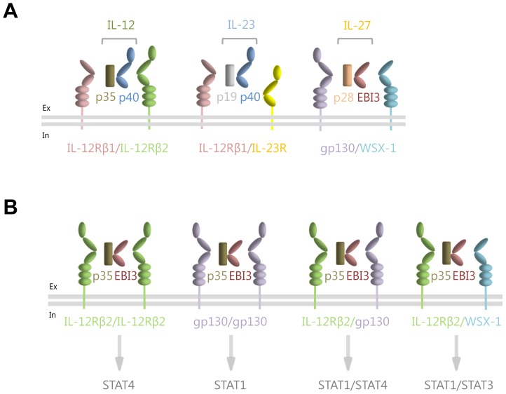
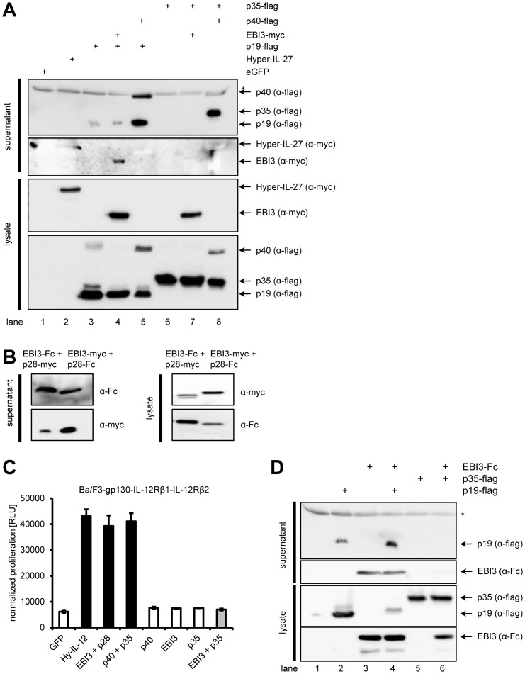
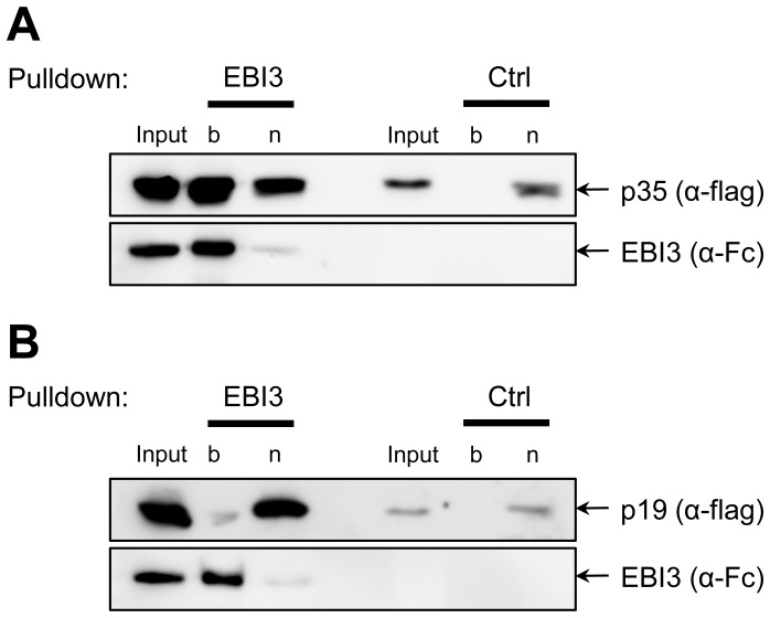
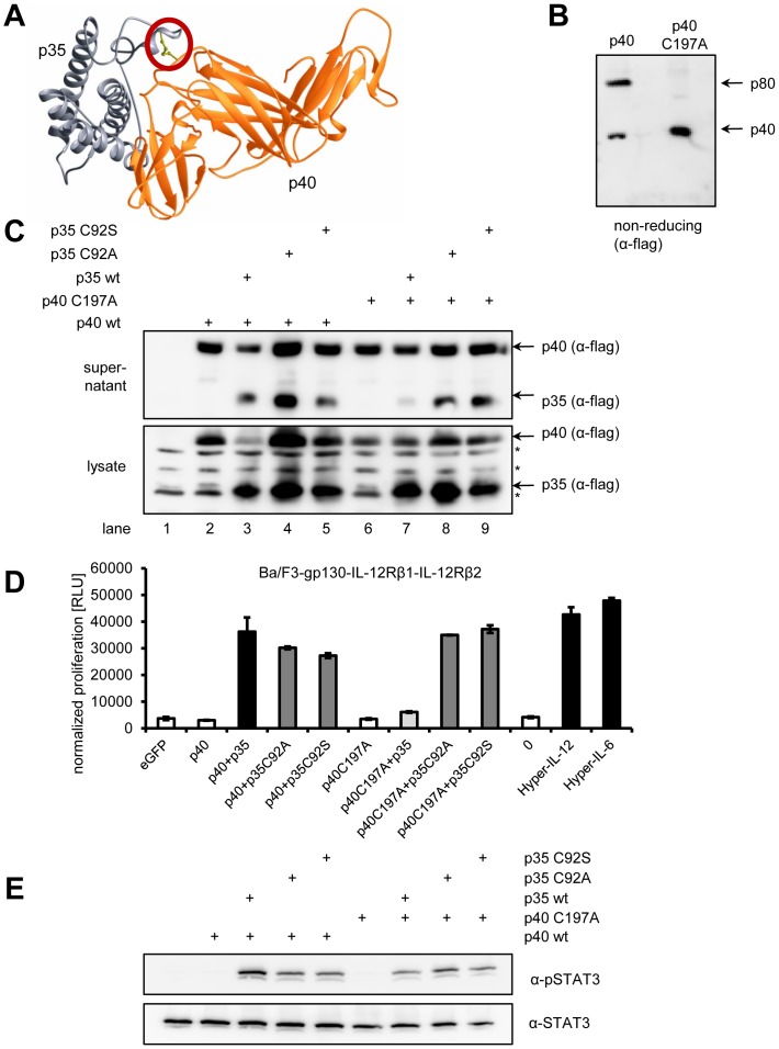
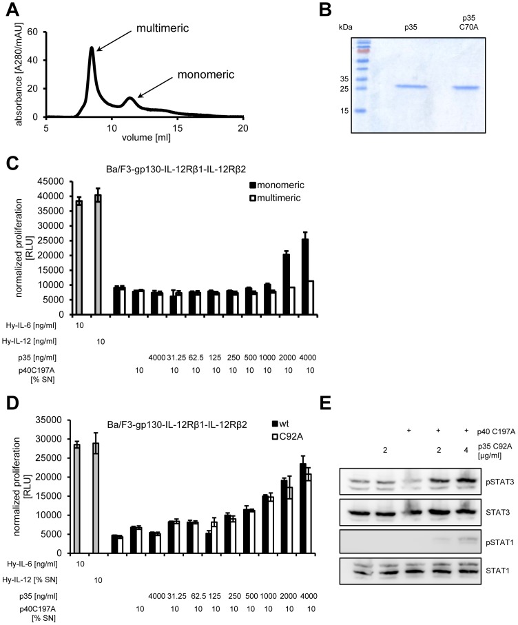
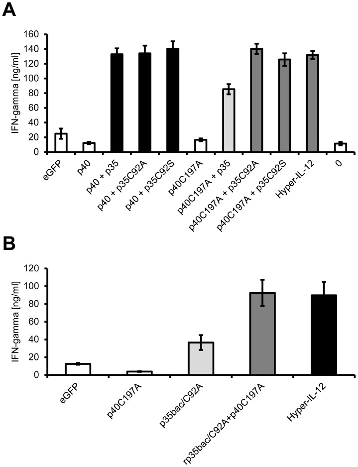
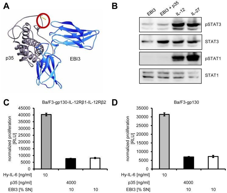
The Interleukin (IL)-12 family contains several heterodimeric composite cytokines which share subunits among each other. IL-12 consists of the subunits p40 (shared with IL-23) and p35. p35 is shared with the composite cytokine IL-35 which comprises of the p35/EBI3 heterodimer (EBI3 shared with IL-27). IL-35 signals via homo- or heterodimers of IL-12Rβ2, gp130 and WSX-1, which are shared with IL-12 and IL-27 receptor complexes, respectively. p35 was efficiently secreted in complex with p40 as IL-12 but not with EBI3 as IL-35 in several transfected cell lines tested which complicates the analysis of IL-35 signal transduction. p35 and p40 but not p35 and EBI3 form an inter-chain disulfide bridge. Mutation of the responsible cysteine residue (p40C197A) reduced IL-12 formation and activity only slightly. Importantly, the p40C197A mutation prevented the formation of antagonistic p40 homodimers which enabled the in vitro reconstitution of biologically active IL-12 with p35 produced in bacteria (p35bac). Reconstitution of IL-35 with p35bac and EBI3 did, however, fail to induce signal transduction in Ba/F3 cells expressing IL-12Rβ2 and gp130. In summary, we describe the in vitro reconstitution of IL-12, but fail to produce recombinant IL-35 by this novel approach.


Conflict of interest statement

Competing Interests: The authors have declared that no competing interests exist.

Figures

Figure 1
Figure 1. Schematic overview of the IL-12 family of cytokines.
(A) Interleukin-12 (consisting of the p35 and the p40 subunits) signals via a heterodimer of the two β-receptors IL-12Rβ1 and IL-12Rβ2. Interleukin-23 (consisting of the p19 and the p40 subunits) signals via a heterodimer of the two β-receptors IL-12Rβ1 and the unique IL-23R. Interleukin-27 (consisting of its subunits p28 and EBI3) engages signaling via gp130 and the unique β-receptor WSX-1. (B) Interleukin-35 (consisting of its subunits p35 and EBI3) is able to signal via four different combinations of β-receptors, resulting in the phosphorylation and thus activation of different STAT proteins. Binding of IL-35 to an IL-12Rβ2 homodimer induces the activation of STAT4 homodimers , binding to a gp130 homodimer activates STAT1 homodimers , binding to an IL-12Rβ2/gp130 heterodimer induces STAT1/STAT4 heterodimerization , and binding of IL-35 to a IL-12Rβ2/WSX-1 heterodimer induces the activation of STAT1 and STAT3 .
Figure 2
Figure 2. In contrast to other Hyper-cytokines, Hyper-IL-35 is not efficiently secreted from cells.
(A) Schematic representation of the six different Hyper-constructs used in this study. In Hyper-IL-6, the extracellular domains of the IL-6R are fused to IL-6, whereas in Hyper-IL-30 p28/IL-30 replaces IL-6. Hyper-IL-27 represents a composite cytokine where EBI3 is fused via a flexible linker to p28/IL-30. Hyper-IL-12 depicts p40 fused to p35, and Hyper-IL-35 is a linker-based fusion-protein of EBI3 and p35. All Hyper-cytokines contain a C-terminal myc-tag with the exception of Hyper-IL-6 and Hyper-IL-30, which are untagged. (B) HEK293 cells were transiently transfected with the different constructs shown in panel (A) or a control plasmid containing eGFP. Supernatant was taken 48 h post transfection. Cells were lysed, and expression and secretion of the different Hyper-cytokines was assessed by Western blotting with monoclonal antibodies against the IL-6R (for the detection of Hyper-IL-6 and Hyper-IL-30) or the myc-tag (for the detection of Hyper-IL-27, Hyper-IL-35, Hyper-IL-35_GGGGS and Hyper-IL-12). Western blots shown are representative of three different experiments with similar outcomes. (C) Equal amounts of Ba/F3-IL-12Rβ1-IL-12Rβ2 cells were incubated with 10% of supernatants derived from HEK293 cells described in panel (B). As a further negative control, cells were incubated without the addition of supernatant (right column). Cellular proliferation was determined 48 h later as described in Materials and Methods . (D) Equal amounts of Ba/F3-IL-12Rβ1-IL-12Rβ2 cells were stimulated with supernatants from HEK293 cells transiently transfected with the constructs indicated above the Western blots for 15 min. Phosphorylation of STAT3 and STAT1 was determined per Western blotting. Total amounts of STAT1 and STAT3 were visualized as internal loading control. The Western blots shown are representative of three different experiments with similar outcomes, and the proliferation assay was measured in triplicates and is representative out two performed experiments.
Figure 3
Figure 3. p28 and EBI3 (IL-27), p40 and p35 (IL-12) as well as p40 and p19 (IL-23) form heterodimeric cytokines and are efficiently secreted, but not EBI3 and p35 (IL-35).
(A) HEK293 cells were transiently transfected with plasmids encoding the constructs indicated above the Western blots. Supernatant was taken 48 h post transfection. Cells were lysed, and expression and secretion of the different cytokines was assessed by Western blotting with monoclonal antibodies against the flag- or the myc-tag. Western blots shown are representative of three different experiments with similar outcomes. The asterisk denotes an unspecific band detected by the flag-antibody. (B) HEK293 cells were transiently transfected with plasmids containing either EBI3-Fc and p28-myc or EBI3-myc and p28-Fc. Expression and secretion of the respective proteins was analyzed by Western blotting as described in panel (A) with antibodies directed against the Fc- or the myc-tag. (C) Equal amounts of Ba/F3-IL-12Rβ1-IL-12Rβ2 cells were incubated with 10% of supernatants derived from transfected cells described in panel (A) and (B). Cellular proliferation was determined 48 h later as described in Materials and Methods . (D) HEK293 cells were transiently transfected with plasmids encoding the constructs indicated above the Western blots. Supernatant was taken 48 h post transfection. Cells were lysed, and expression and secretion of the different cytokines was assessed by Western blotting with monoclonal antibodies against the flag- or the Fc-tag. Western blots shown are representative of three different experiments with similar outcomes. The asterisk denotes an unspecific band detected by the flag-antibody.
Figure 4
Figure 4. Interaction between p35 and EBI3.
(A) HEK293 cells were transiently transfected with either p35 or EBI3. Cells were lysed, and lysates were mixed. To check for unspecific binding of p35 to the beads, lysates of p35-transfected cells without EBI were used as control (Ctrl). Pulldown was performed as described in Materials and Methods . Proteins were analyzed by Western blotting with antibodies against flag- and Fc-tag. (B) The experiment was performed as described under panel (A), but p19-transfected cells were used instead of p35. “b” denotes the bound fraction, “n” the non-bound proteins.
Figure 5
Figure 5. The disulfide bond p35C92-C197p40 is dispensable for the biological activity of IL-12.
(A) Schematic representation of IL-12 comprising p35 (gray) and p40 (orange) according to . The inter-molecular disulfide bond p35C92-C197p40 is highlighted with a red circle. (B) 20 µl conditioned supernatant of HEK293 cells transiently transfected with either p40 wildtype or p40C197A were separated by SDS-PAGE under non-reducing conditions and proteins visualized by Western blotting with a flag-specific antibody. (C) HEK293 cells were transiently transfected with plasmids encoding the constructs indicated above the Western blots. Supernatant was harvested 48 h post transfection. Cells were lysed, and expression and secretion of the different cytokines was assessed by Western blotting with monoclonal antibodies against the flag-tag. Unspecific bands detected by the flag-antibody are denoted with asterisks. (D) Ba/F3-IL-12Rβ1-IL-12Rβ2 cells were incubated with 10% of supernatants derived from HEK293 cells transiently transfected with the constructs given below the diagram. Cellular proliferation was determined 48 h later as described in Materials and Methods . (E) Equal amounts of Ba/F3-IL-12Rβ1-IL-12Rβ2 cells were stimulated with supernatants from HEK293 cells transiently transfected with the constructs indicated above the Western blots for 15 min. Phosphorylation of STAT3 was determined per Western blotting. Total amounts of STAT3 were visualized as internal loading control. The Western blots shown are representative of three different experiments with similar outcomes, and the proliferation assay was measured in triplicates and is representative of two performed experiments.
Figure 6
Figure 6. Recombinant p35 from bacteria forms biologically active IL-12.
(A) Refolded p35bac and p35bac/C92A were subjected to size exclusion chromatography. Monomeric protein was separated from multimeric assemblies on a Superdex 75 10/300 GL equilibrated in 50 mM Tris-HCl (pH 8.0) containing 250 mM NaCl. (B) Purity of the bacterial produced p35bac and p35bac/C92A was analyzed by SDS-PAGE on a reducing gel via Coomassie brilliant blue staining. 5 µg protein were loaded per lane. (C) Equal amounts of Ba/F3-IL-12Rβ1-IL-12Rβ2 cells were incubated with 10 ng/ml Hyper-IL-6, 10% conditioned supernatant containing Hyper-IL-12, or 10% conditioned supernatant containing p40C197A with increasing amounts of either monomeric or multimeric p35. (D) Equal amounts of Ba/F3-IL-12Rβ1-IL-12Rβ2 cells were treated as described under panel (C), but with either recombinant p35bac or p35bac/C92A (0–4000 ng/ml). Cellular proliferation in both experiments was determined 48 h later as described in Materials and Methods . (E) Equal amounts of Ba/F3-gp130-IL-12Rβ1-IL-12Rβ2 cells were incubated with 50% conditioned supernatant containing p40C197A with or without 2 or 4 µg/ml p35bac/C92A for 15 min. Phosphorylation of STAT1 and STAT3 was determined per Western blotting. Total amounts of STAT1 and STAT3 were visualized as internal loading control. The data shown are representative of two different experiments with similar outcomes.
Figure 7
Figure 7. IL-12 induces the expression of Interferon-γ in murine T cells.
(A) Murine CD4+ T cells were enriched by positive selection from single cell suspended splenocytes and lymphnode cells. 1×10⁵ cells per well were cultured on plates coated with 0.5 µg/ml anti-CD3 and 2 µg/ml soluble anti-CD28. 10% of the indicated cell culture supernatant was added and supernatants were harvested after three days. Secretion of IFN-γ was determined via ELISA. (B) The experiment was performed as described under panel (A). Where indicated, 2 µg/ml p35bac/C92A were added. The experiments were performed with at least three individual mice, and data from one representative animal are shown.
Figure 8
Figure 8. IL-35, formed by recombinant p35, does not induce proliferation or STAT phosphorylation in Ba/F3-gp130-IL-12Rβ1-IL-12Rβ2 cells.
(A) Schematic representation of IL-35 comprising of p35 (gray) and EBI3 (blue). C92 of p35, which is not connected via a disulfide bond with EBI3, is highlighted with a red circle. (B) Equal amounts of Ba/F3-gp130-IL-12Rβ1-IL-12Rβ2 cells were incubated with 50% conditioned supernatant containing EBI3 with or without 2 µg/ml p35bac/C92A for 15 min. Phosphorylation of STAT1 and STAT3 was determined per Western blotting. Total amounts of STAT1 and STAT3 were visualized as internal loading control. (C) Equal amounts of Ba/F3-gp130-IL-12Rβ1-IL-12Rβ2 cells were incubated with 10 ng/ml Hyper-IL-6 or 10% conditioned supernatant containing EBI3, either with or without 4 µg/ml recombinant p35bac. Cellular proliferation was determined 48 h later as described in Materials and Methods . (D) The experiment was performed as described under panel (C), but with Ba/F3-gp130 instead of Ba/F3-gp130-IL-12Rβ1-IL-12Rβ2 cells. Cellular proliferation was determined 48 h later as described in Materials and Methods . The Western blots shown as well as the proliferation assays are representative of three different experiments with similar outcomes.

References

    1. Garbers C, Hermanns H, Schaper F, Müller-Newen G, Grötzinger J, et al. (2012) Plasticity and cross-talk of Interleukin 6-type cytokines. Cytokine Growth Factor Rev 23: 85–182. - PubMed
    1. Jones L, Chaturvedi V, Uyttenhove C, Van Snick J, Vignali DA (2012) Distinct subunit pairing criteria within the heterodimeric IL-12 cytokine family. Mol Immunol 51: 234–244. - PMC - PubMed
    1. Hölscher C (2004) The power of combinatorial immunology: IL-12 and IL-12-related dimeric cytokines in infectious diseases. Med Microbiol Immunol 193: 1–17. - PubMed
    1. Gillessen S, Carvajal D, Ling P, Podlaski F, Stremlo D, et al. (1995) Mouse interleukin-12 (IL-12) p40 homodimer: a potent IL-12 antagonist. Eur J Immunol 25: 200–206. - PubMed
    1. Ling P, Gately M, Gubler U, Stern A, Lin P, et al. (1995) Human IL-12 p40 homodimer binds to the IL-12 receptor but does not mediate biologic activity. J Immunol 154: 116–127. - PubMed

Publication types

MeSH terms