. 2014 Sep 26;9(9):e108746.
doi: 10.1371/journal.pone.0108746. eCollection 2014.

Transovum transmission of trypanosomatid cysts in the Milkweed bug, Oncopeltus fasciatus

Affiliations

Transovum transmission of trypanosomatid cysts in the Milkweed bug, Oncopeltus fasciatus

Felipe de Almeida Dias et al. PLoS One. .

Abstract

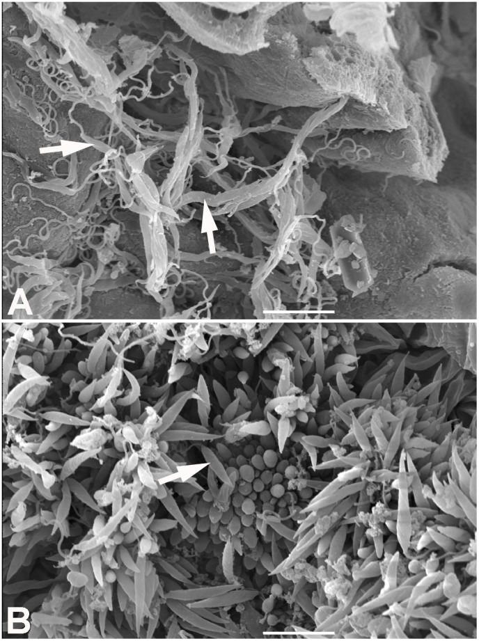
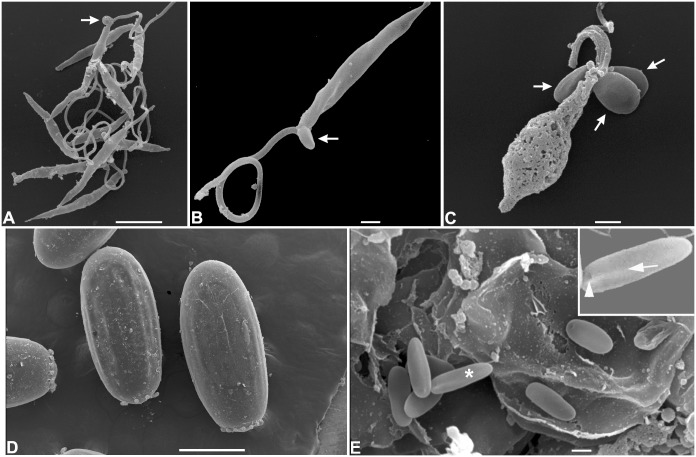
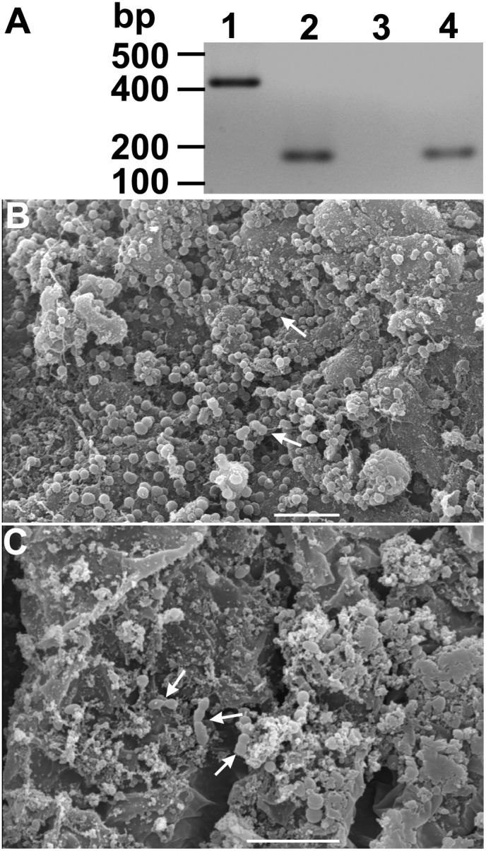
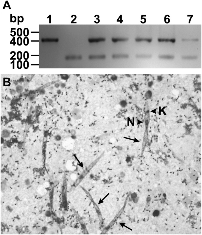
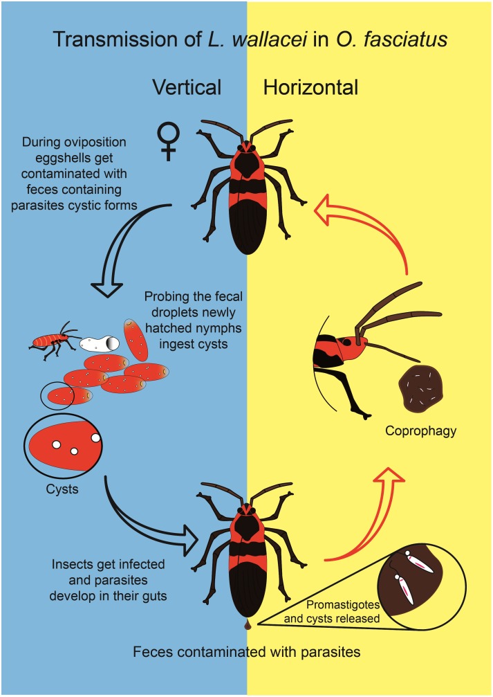
Leptomonas wallacei is a trypanosomatid that develops promastigotes and cystic forms in the gut of the hemipteran insect Oncopeltus fasciatus. Insect trypanosomatids are thought to be solely transmitted from one host to another through the ingestion of parasite-contaminated feces. However, here we show that L. wallacei cysts present on the eggshells of eggs laid by O. fasciatus can also act as infective forms that are transmitted to the insect offspring. Newly hatched O. faciatus nymphs are parasite-free, but some of them become contaminated with L. wallacei after feeding on eggshell remnants. The present study is the first report of transovum transmission of a trypanosomatid, a process that may have a relevant role in parasite's within-host population dynamics.


Conflict of interest statement

Competing Interests: The authors have declared that no competing interests exist.

Figures

Figure 1
Figure 1. Scanning electron microscopy of Oncopeltus fasciatus guts infected with Leptomonas wallacei.
(A) Midgut of L. wallacei-infected O. fasciatus. The arrows indicate large numbers of parasites near the midgut wall. (B) Hindgut of L. wallacei-infected O. fasciatus. The image shows a massive presence of flagellates. Most of those are attached to the intestinal wall of the hindgut by their flagella, so only their slender bodies can be seen. One of the short-sized flagellates can be seen in the lumen (arrow). Bars = 10 µm.
Figure 2
Figure 2. Scanning electron micrograph of feces and eggs of Oncopeltus fasciatus infected with Leptomonas wallacei.
(A) Promastigote forms of L. wallacei in fresh feces showing typical characteristics of live cells and cystic forms (arrow). Bar = 5 µm. (B) High magnification of a promastigote form showing one cystic form (arrow) near to its flagella. Bar = 1 µm. (C) Promastigote form present in the naturally dried feces showing extensive membrane damage and three cystic forms. Note that the cystic forms (arrows) do not show any surface damage. Bar = 1 µm. (D) Low magnification of the surface of the eggs. Bar = 500 µm. (E) High magnification of egg surface showing typical cystic forms of L. wallacei (arrows). Bar = 1 µm.
Figure 3
Figure 3. Oncopeltus fasciatus hatched from eggs submitted to surface asepsis were Leptomonas wallacei-free.
(A) Representative gel electrophoresis of PCR-amplified DNA samples extracted from eggs and whole insect guts. Lane 1- The DNA extracted from an axenic culture of L. wallacei was amplified with primers specific for parasite detection. Lane 2- Sample of pooled DNA extracted from eggs collected at the infected colony and submitted to surface asepsis was concomitantly amplified with primers specific for parasite or insect DNA detection. Lanes 3 and 4- Sample of pooled DNA, extracted from 3 pools of five insect guts of insects hatched from eggs submitted to asepsis, was PCR-amplified with primers specific for parasite (lane 3) or insect DNA (lane 4) detection, respectively. On the left, the positions of molecular size markers are shown in base pairs. The figure represents a negative image of the gel. (B and C) Scanning electron microscopy of O. fasciatus midgut and hindgut, respectively. The micrographs show the presence of bacteria (arrows) but absence of parasites. Bars = 10 µm.
Figure 4
Figure 4. Detection of parasites after experimental transovum transmission of Leptomonas wallacei parasites by Oncopeltus fasciatus.
(A) Detection of parasite infection by PCR. Lane 1- The DNA extracted from an axenic culture of L. wallacei was amplified with primers specific for parasite detection. Lane 2- A pool of DNA samples extracted from gut of parasite-free insects was concomitantly amplified with primers specific for parasite and insect DNA detection. Lanes 3–7- Representative DNA samples extracted from guts of insects that fed on sunflower seeds contaminated with eggshels collected from infected colony were concomitantly amplified with primers specific for parasite and insect DNA detection. On the left, the positions of molecular size markers are shown in base pairs. The figure represents a negative image of the gel. (B) Detection of parasite infection by optical microscopy. The representative micrograph shows Giemsa-stained parasites (arrows) in the gut contents of newly infected insects. The arrowheads indicate the nucleus (N) and kinetoplast (K) of the parasite. Magnification = 400 x.
Figure 5
Figure 5. Horizontal and vertical transmission of Leptomonas wallacei by Oncopeltus fasciatus.
The insects get infected by feeding on feces contaminated with parasites. L. wallacei produces cystic forms, which are present in the insect guts; the feces contaminate the eggshells. Newly hatched nymphs feed on egg remnants and acquire infection. The cartoon represents a model of transmission based upon previous data and the results shown here.

References

    1. Rodrigues JC, Godinho JL, de Souza W (2014) Biology of human pathogenic trypanosomatids: epidemiology, lifecycle and ultrastructure. Subcell Biochem 74: 1–42. - PubMed
    1. Simpson AG, Stevens JR, Lukes J (2006) The evolution and diversity of kinetoplastid flagellates. Trends Parasitol 22: 168–174. - PubMed
    1. Fokin SI, Schrallhammer M, Chiellini C, Verni F, Petroni G (2014) Free-living ciliates as potential reservoirs for eukaryotic parasites: occurrence of a trypanosomatid in the macronucleus of Euplotes encysticus . Parasit Vectors 7: 203. - PMC - PubMed
    1. Podlipaev S (2001) The more insect trypanosomatids under study-the more diverse Trypanosomatidae appears. Int J Parasitol 31: 648–652. - PubMed
    1. McGhee RB, Hanson WL (1964) Comparison of the life cycle of Leptomonas oncopelti and Phytomonas elmassiani . J Protozool 11: 555–562. - PubMed

Publication types

LinkOut - more resources