. 2014 Dec;122(12):1306-13.
doi: 10.1289/ehp.1408453. Epub 2014 Sep 26.

Structural and functional profiling of environmental ligands for estrogen receptors

Affiliations

Structural and functional profiling of environmental ligands for estrogen receptors

Vanessa Delfosse et al. Environ Health Perspect. 2014 Dec.

Abstract

Background: Individuals are exposed daily to environmental pollutants that may act as endocrine-disrupting chemicals (EDCs), causing a range of developmental, reproductive, metabolic, or neoplastic diseases. With their mostly hydrophobic pocket that serves as a docking site for endogenous and exogenous ligands, nuclear receptors (NRs) can be primary targets of small molecule environmental contaminants. However, most of these compounds are chemically unrelated to natural hormones, so their binding modes and associated hormonal activities are hardly predictable.

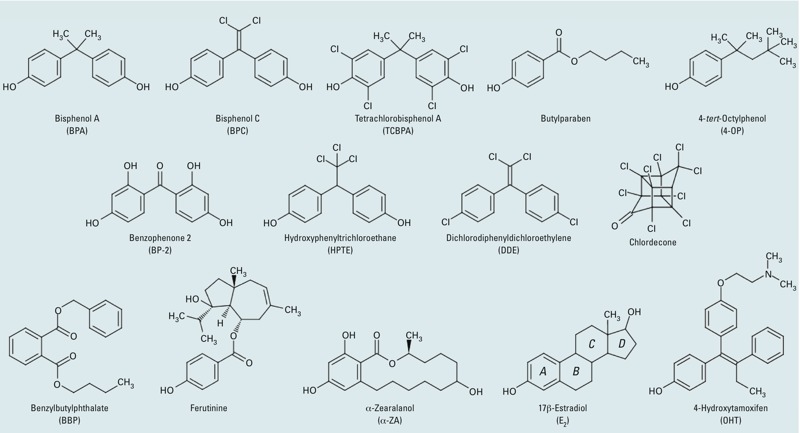
Objectives: We conducted a correlative analysis of structural and functional data to gain insight into the mechanisms by which 12 members of representative families of pollutants bind to and activate the estrogen receptors ERα and ERβ.

Methods: We used a battery of biochemical, structural, biophysical, and cell-based approaches to characterize the interaction between ERs and their environmental ligands.

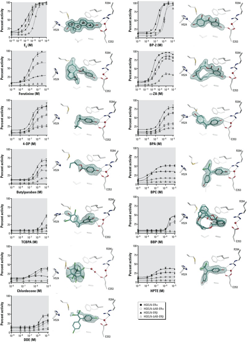
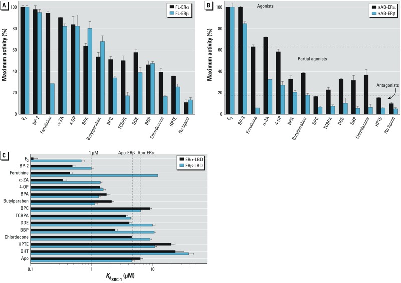
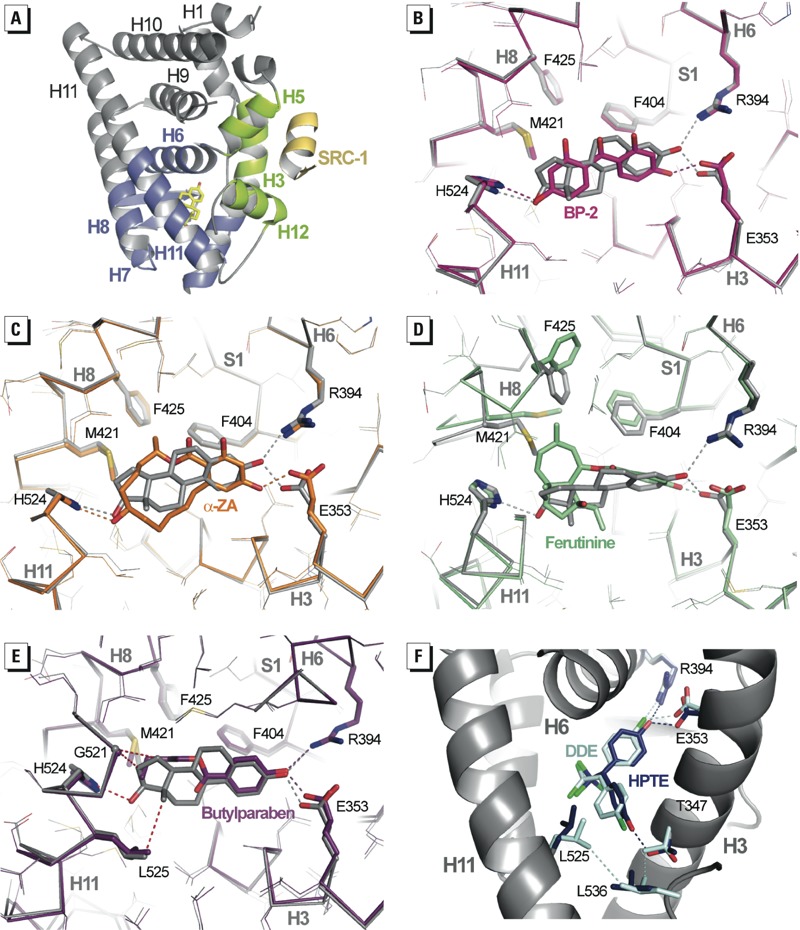
Results: Our study revealed that the chemically diverse compounds bound to ERs via varied sets of protein-ligand interactions, reflecting their differential activities, binding affinities, and specificities. We observed xenoestrogens binding to both ERs-with affinities ranging from subnanomolar to micromolar values-and acting in a subtype-dependent fashion as full agonists or partial agonists/antagonists by using different combinations of the activation functions 1 and 2 of ERα and ERβ.

Conclusions: The precise characterization of the interactions between major environmental pollutants and two of their primary biological targets provides rational guidelines for the design of safer chemicals, and will increase the accuracy and usefulness of structure-based computational methods, allowing for activity prediction of chemicals in risk assessment.


Conflict of interest statement

The authors declare they have no actual or potential competing financial interests.

Figures

Figure 1
Figure 1
Chemical structures of the natural agonist E2, the synthetic antagonist OHT, and the various environmental ER ligands used in the present study.
Figure 2
Figure 2
The relative activity of xenoestrogens relies on their different binding modes. (Left) Dose–response curves corresponding to the HGELN-ERα, -∆AB-ERα, -ERβ, and -∆AB-ERβ, luciferase assays of E2 and xenoestrogens. The maximal activity (100%) was obtained with 10 nM E2; values are mean ± SD from three separate experiments. (Right) The interaction networks of E2 and xenoestrogens with LBD residues of ERα. Key for structures: red, oxygen; blue, nitrogen; cyan, carbon; yellow, sulfur; green, chlorine; black dashed lines, hydrogen bonds; red spheres, water molecules. The electron density represents a Fo–Fc simulated annealing omit map contoured at 3σ.
Figure 3
Figure 3
Differential involvement of AFs in ERs. HGELN-ERs cells (A) and HGELN-∆AB-ERs cells (B) were incubated with 10 nM E2 or 10 μM ER exogenous ligands. The maximal luciferase activity (100%) was obtained with 10 nM E2; values are the mean ± SD from three separate experiments. (In B, the horizontal dotted lines highlight the partition of the ligands into three classes based on fluorescence anisotropy data. (C) Fluorescence anisotropy data showing the relative affinity of the SRC-1 NR2 peptide for ERα LBD or ERβ LBD in the absence of ligand or in the presence of saturating concentrations of E2 or xenoestrogens. Ligands are classified as agonists (Kd ≤ 1 μM), partial agonists (1 μM ≤ Kd ≤ values for apo-ERα or -ERβ), or antagonists (Kd ≥ values for apo-ERα or -ERβ).
Figure 4
Figure 4
Xenoestrogens use diverse binding modes. (A) The entire structure of the ERα LBD in complex with E2 and SRC-1 coactivator peptide (yellow); the structure shows the AF‑2 surface formed by helices H3, H5, and H12 (green), and the lower part of the LBD (blue) encloses the ligand-binding pocket (LBP). (B–E) Interaction networks of BP-2 (B; pink), α-ZA (C; orange), ferutinine (D; green), and butylparaben (E; purple) with residues of the LBP compared with that of E2 (gray). In (E), red dashed lines represent the interactions lost in the butylparaben complex structure. (F) HPTE and DDE adopt the orientation previously observed for BPC allowing HPTE to interact with residue T347. This position results in the disruption of the hydrophobic network involving helices H3 and H11 and the loop preceding H12, thereby destabilizing the AF‑2 surface. Color code: red, oxygen; blue, nitrogen; cyan, carbon; yellow, sulfur; green, chlorine; dashed lines, hydrogen bonds and hydrophobic interactions.
Figure 5
Figure 5
Methionine 421 confers plasticity and adaptability to ERα LBP. Structure superposition of E2-bound ERβ LBD (yellow) with (A) ferutinine-bound ERα LBD (green), or (B) BBP-bound ERα LBD (gray). The presence of I373 in ERβ instead of M421 in ERα will induce a shift of bulky ligands toward helix H12 thus lowering the stability of the AF‑2.

Comment in

References

    1. Bossard C, Busson M, Vindrieux D, Gaudin F, Machelon V, Brigitte M, et al. 2012Potential role of estrogen receptor beta as a tumor suppressor of epithelial ovarian cancer. PloS one 79e44787; 10.1371/journal.pone.0044787 - DOI - PMC - PubMed
    1. Bruning JB, Parent AA, Gil G, Zhao M, Nowak J, Pace MC, et al. Coupling of receptor conformation and ligand orientation determine graded activity. Nat Chem Biol. 2010;6(11):837–843. - PMC - PubMed
    1. Couse JF, Korach KS. Estrogen receptor null mice: what have we learned and where will they lead us? Endocr Rev. 1999;20(3):358–417. - PubMed
    1. Curtis SW, Washburn T, Sewall C, DiAugustine R, Lindzey J, Couse JF, et al. Physiological coupling of growth factor and steroid receptor signaling pathways: estrogen receptor knockout mice lack estrogen-like response to epidermal growth factor. Proc Natl Acad Sci USA. 1996;93(22):12626–12630. - PMC - PubMed
    1. De Coster S, van Larebeke N.2012Endocrine-disrupting chemicals: associated disorders and mechanisms of action. J Environ Public Health 2012713696; 10.1155/2012/713696 - DOI - PMC - PubMed

Publication types

MeSH terms