. 2015 May 1:215:106-16.
doi: 10.1016/j.ygcen.2014.09.010. Epub 2014 Sep 26.

Structural libraries of protein models for multiple species to understand evolution of the renin-angiotensin system

Affiliations

Structural libraries of protein models for multiple species to understand evolution of the renin-angiotensin system

Jeremy W Prokop et al. Gen Comp Endocrinol. .

Abstract

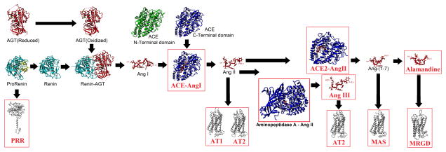
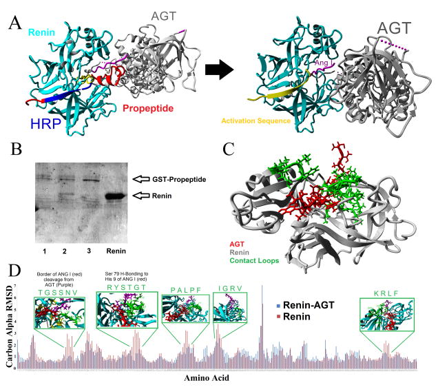
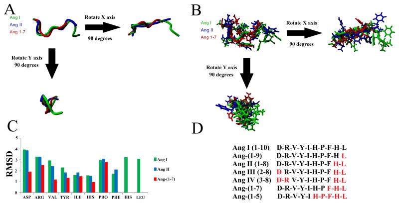
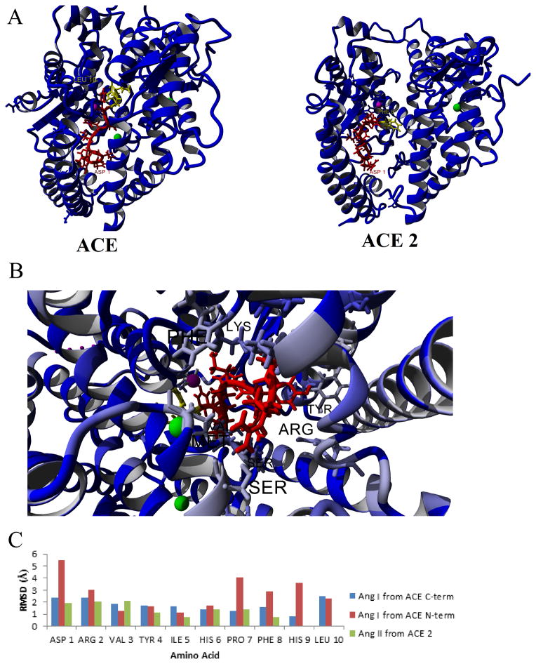
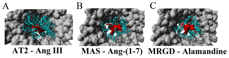
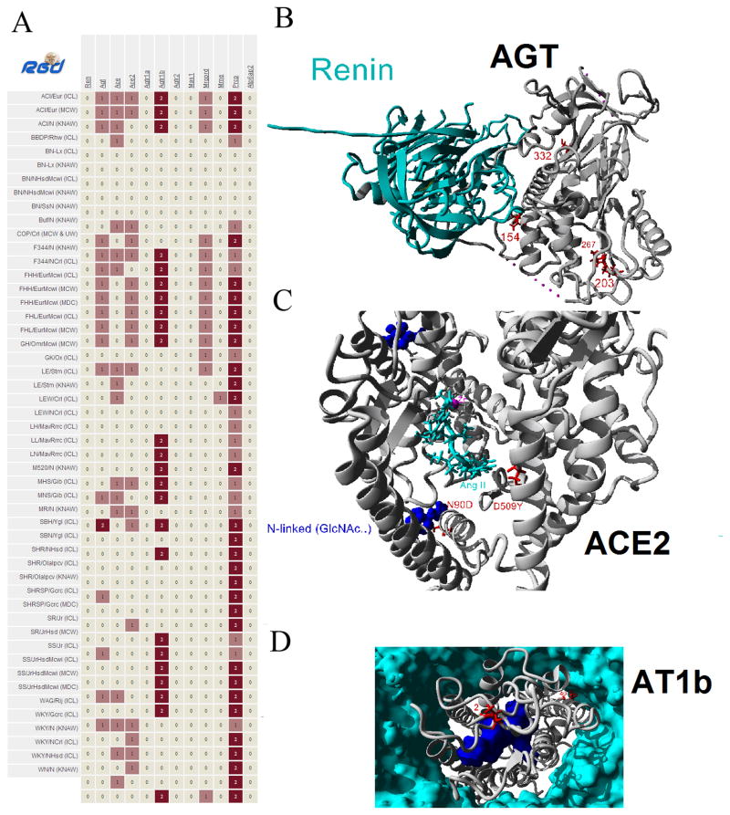
The details of protein pathways at a structural level provides a bridge between genetics/molecular biology and physiology. The renin-angiotensin system is involved in many physiological pathways with informative structural details in multiple components. Few studies have been performed assessing structural knowledge across the system. This assessment allows use of bioinformatics tools to fill in missing structural voids. In this paper we detail known structures of the renin-angiotensin system and use computational approaches to estimate and model components that do not have their protein structures defined. With the subsequent large library of protein structures, we then created a species specific protein library for human, mouse, rat, bovine, zebrafish, and chicken for the system. The rat structural system allowed for rapid screening of genetic variants from 51 commonly used rat strains, identifying amino acid variants in angiotensinogen, ACE2, and AT1b that are in contact positions with other macromolecules. We believe the structural map will be of value for other researchers to understand their experimental data in the context of an environment for multiple proteins, providing pdb files of proteins for the renin-angiotensin system in six species. With detailed structural descriptions of each protein, it is easier to assess a species for use in translating human diseases with animal models. Additionally, as whole genome sequencing continues to decrease in cost, tools such as molecular modeling will gain use as an initial step in designing efficient hypothesis driven research, addressing potential functional outcomes of genetic variants with precompiled protein libraries aiding in rapid characterizations.

Keywords: Angiotensin peptides; Comparative modeling; Rat genetics; Renin-angiotensin system; Sequence-to-structure-to-function analysis.


Figures

Figure 1
Figure 1. Multiple components of the renin-angiotensin system
Structural analysis of the RAS with components that are characterized using the in silico approaches in this paper boxed in red. Biochemically determined structures are known for AGT (reduced/oxidized), Prorenin, Renin, Renin-AGT, Ang I, Ang II, Ang-(1-7), ACE (N/C-terminal domains), and ACE 2. Models were used for the PRR, AT1, AT2, MAS, MRGD, ACE-Ang I, Aminopeptidase AAng II, and ACE2–Ang II. In addition to the models shown here we have generated potential structures for Ang- Neprilysin and Ang-PRCP.
Figure 2
Figure 2. Role of cleavage of the propeptide in activation of Renin and binding of AGT
A) Addition of the propeptide (red) of Prorenin results in the activation sequence (yellow) to cover the active site as well as steric hindrance with the contact points between Renin and AGT. Cleavage of the propeptide results in the movement of the activation domain, allowing for Ang I (magenta) proper cleavage and additional contact points between Renin and AGT. The proposed potential therapeutic RAS peptide, known as the handle region peptide (HRP, blue), is identified to contribute to the beta sheet packing that allosterically modifies the activation sequence. B) Pull down experiments of Renin with the GST-Propeptide on glutathione sepharose. Sample 1 is a control of GST-Propeptide pull down, 2 is the pull down with additional Renin added in for co-pull down, 3 has Renin and the shorter ProR peptide (longer version of the human HRP) added resulting in a loss of Renin co-pull down, and the final lane contains the purified Renin protein. C) Interaction between a segment of the N-terminus of AGT (red) in the active site of Renin (gray) used for molecular dynamics simulations. D) Molecular dynamics simulation comparing renin alone (red) or complexed with the AGT fragment (blue) showing the carbon alpha RMSD differences for each amino acid on Renin. For areas with large differences in dynamics, shown above with a green box are the amino acids contacts with AGT in the loops of Renin which are highlighted in green on the structure of Renin in C.
Figure 3
Figure 3. Known and modeled structures of the Ang peptides
A–B) Structural alignment of Ang I (green), Ang II (blue), and Ang-(1-7) (red) aligned together showing just the backbone (A) or the side chains (B). C) Molecular dynamic simulations on each peptide for 10 nanoseconds showing the average movement of the carbon alpha for each of the amino acids of the peptide. D) Sequences of the various Ang peptides used for Autodock experiments. Amino acids in red were removed from Ang I to produce the peptide sequence shown in black.
Figure 4
Figure 4. Docking of Ang peptides to ACE and ACE2
A) Structures of the top docking of Ang I to ACE (left) or Ang II to ACE 2 (right). The Ang peptides are shown in red with the amino acids cleaved off in yellow. The Zn active site is magenta and the Cl ions are green. B) Variant amino acids (grey to light blue) between the N and Cterminus showing contributing amino acids to variant binding of Ang I. C) Molecular dynamics simulations showing the movement of Ang I to either the C-terminus (blue) or the N-terminus (red) of ACE or Ang II to ACE 2 (green).
Figure 5
Figure 5. Docking of Ang peptides into various uncharacterized receptors
A) The AT2 receptor model (cyan) interacting with Ang III (red) in a lipid membrane (gray). B) The MAS (cyan) model in a lipid membrane (gray) with Ang 1-7 (red) docked into the binding site as determined by the best conformation in 100 Autodock predictions. C) Model of MRGD (cyan) with Alamandine (red) bound into the binding pocket based on the docking results of MAS/Ang-(1-7).
Figure 6
Figure 6. Variation of RAS proteins in the multiple rat genomes
A) Nonsynonymous mutations identified in the multiple rat genomes (left) for several of the RAS genes (top). B) Based on structural assessment of all rat variants, the amino acid 154 (red) variant of AGT (gray) is in close proximity to contacts with Renin (cyan). C) Amino acid 90 (red) variant of ACE2 (gray) is a cite of known site for N-linked glycosylation (blue) while amino acid 509 variant is located close to Ang II (cyan) binding. D) Amino acid 40 (red) variant in AT1b (gray) is located at a membrane (cyan) contact point on a helix with known contacts with the Ang II peptide (blue).

References

    1. Adzhubei IA, Schmidt S, Peshkin L, Ramensky VE, Gerasimova A, Bork P, Kondrashov AS, Sunyaev SR. A method and server for predicting damaging missense mutations. Nat Methods. 2010;7:248–249. doi: 10.1038/nmeth0410-248. - DOI - PMC - PubMed
    1. Ager EI, Neo J, Christophi C. The renin-angiotensin system and malignancy. Carcinogenesis. 2008;29:1675–1684. doi: 10.1093/carcin/bgn171. - DOI - PubMed
    1. Atanur SS, Diaz AG, Maratou K, Sarkis A, Rotival M, Game L, Tschannen MR, Kaisaki PJ, Otto GW, Ma MCJ, Keane TM, Hummel O, Saar K, Chen W, Guryev V, Gopalakrishnan K, Garrett MR, Joe B, Citterio L, Bianchi G, McBride M, Dominiczak A, Adams DJ, Serikawa T, Flicek P, Cuppen E, Hubner N, Petretto E, Gauguier D, Kwitek A, Jacob H, Aitman TJ. Genome Sequencing Reveals Loci under Artificial Selection that Underlie Disease Phenotypes in the Laboratory Rat. Cell. 2013;154:691–703. doi: 10.1016/j.cell.2013.06.040. - DOI - PMC - PubMed
    1. Batenburg WW, van den Heuvel M, van Esch J, van Veghel R, Garrelds I, Leijten F, Danser A. The (pro)renin receptor blocker handle region peptide upregulates endothelium-derived contractile factors in aliskiren-treated diabetic transgenic (mREN2)27 rats. J Hypertens Febr 2013. 2013;31:292–302. doi: 10.1097/HJH.0b013e32835c1789. - DOI - PubMed
    1. Bosnyak S, Jones ES, Christopoulos A, Aguilar MI, Thomas WG, Widdop RE. Relative affinity of angiotensin peptides and novel ligands at AT1 and AT2 receptors. Clin Sci Lond Engl 1979. 2011;121:297–303. doi: 10.1042/CS20110036. - DOI - PubMed

Publication types

LinkOut - more resources