Case Reports
. 2015 Jan;12(1):211-9.
doi: 10.1016/j.hrthm.2014.09.051. Epub 2014 Sep 28.

Novel Timothy syndrome mutation leading to increase in CACNA1C window current

Affiliations
Case Reports

Novel Timothy syndrome mutation leading to increase in CACNA1C window current

Nicole J Boczek et al. Heart Rhythm. 2015 Jan.

Abstract

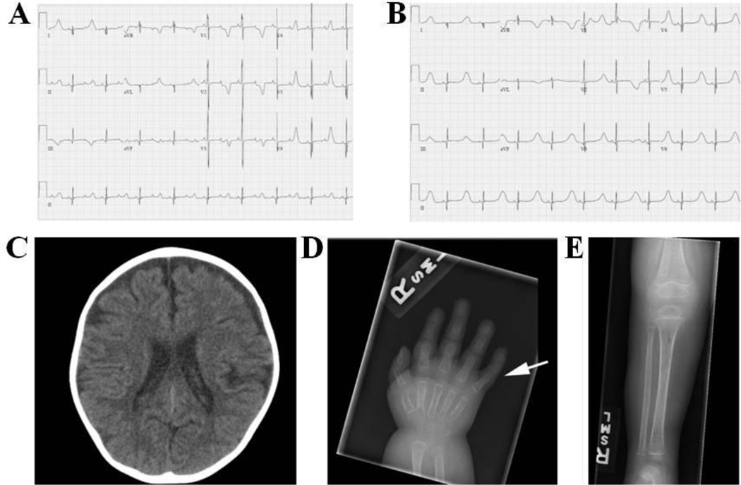
Background: Timothy syndrome (TS) is a rare multisystem genetic disorder characterized by a myriad of abnormalities, including QT prolongation, syndactyly, and neurologic symptoms. The predominant genetic causes are recurrent de novo missense mutations in exon 8/8A of the CACNA1C-encoded L-type calcium channel; however, some cases remain genetically elusive.

Objective: The purpose of this study was to identify the genetic cause of TS in a patient who did not harbor a CACNA1C mutation in exon 8/A, and was negative for all other plausible genetic substrates.

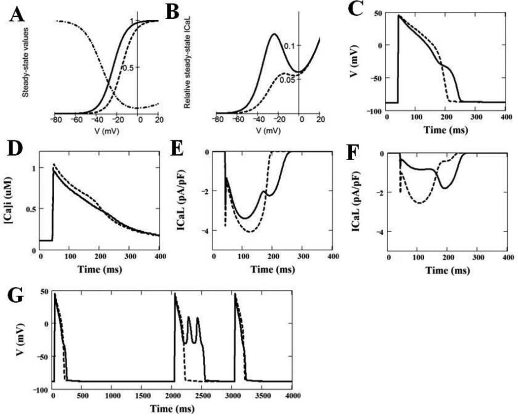
Methods: Diagnostic exome sequencing was used to identify the genetic substrate responsible for our case of TS. The identified mutation was characterized using whole-cell patch-clamp technique, and the results of these analyses were modeled using a modified Luo-Rudy dynamic model to determine the effects on the cardiac action potential.

Results: Whole exome sequencing revealed a novel CACNA1C mutation, p.Ile1166Thr, in a young male with diagnosed TS. Functional electrophysiologic analysis identified a novel mechanism of TS-mediated disease, with an overall loss of current density and a gain-of-function shift in activation, leading to an increased window current. Modeling studies of this variant predicted prolongation of the action potential as well as the development of spontaneous early afterdepolarizations.

Conclusion: Through expanded whole exome sequencing, we identified a novel genetic substrate for TS, p.Ile1166Thr-CACNA1C. Electrophysiologic experiments combined with modeling studies have identified a novel TS mechanism through increased window current. Therefore, expanded genetic testing in cases of TS to the entire CACNA1C coding region, if initial targeted testing is negative, may be warranted.

Keywords: CACNA1C; Genetics; Timothy syndrome; Whole exome sequencing; Window current.


Conflict of interest statement

Conflicts of Interest:

The other authors have no conflicts of interest relevant to this article to disclose.

Figures

Figure 1
Figure 1. Electrocardiograms, CT Scan, and X-Ray Images from the Patient Harboring p.Ile1166Thr-CACNA1C
Electrocardiograms representing 2:1 block at birth (A) and t-wave alternans (B). (C) Non-contrast CT of the head at 3 years 8 months of age showing mild diffuse cerebral parenchymal volume loss. (D) Right hand film at 10 months of age showing 5th finger clinodactyly (arrow). (E) AP tibia-fibula film at 10 months of age demonstrating osteopenia with a permeative appearance of the bone.
Figure 2
Figure 2. CACNA1C Protein Topology
Topology diagram of the CACNA1C channel α-subunit in the membrane. Each repeat is demarcated at the top of the figure. Red circles represent the canonical TS-associated mutations in exon 8 and 8A. The yellow circle represents the p.Ile1166Thr mutation identified within our patient. The blue circle represents the TS mutation identified by Gillis and colleagues.
Figure 3
Figure 3. CACNA1C-Ile1166Thr Reduced ICaL Current in Heterologous Cells
(A) Whole cell ICaL current representative tracings from HEK293 cells expressing CACNA1C-WT or CACNA1C-Ile1166Thr determined from a holding potential of −90 mV to testing potential of +70 mV in 10 mV increments with 500 ms duration. (B) Current-voltage relationship for CACNA1C-WT (n=11) and –Ile1166Thr missense mutation (n=11). All values represent mean SEM. *p<0.05 vs. CACNA1C-WT.
Figure 4
Figure 4. CACNA1C-Ile1166Thr Negatively Shifted ICaL V1/2 in Activation
(A) Activation curves of ICaL CACNA1C-WT (n=11) and –Ile1166Thr mutation (n=11). G/Gmax represents normalized conductance fitted with a Boltzmann function. (B) Inactivation curves of ICaL CACNA1C-WT (n=10) and Ile1166Thr variant (n=4), determined from a holding potential of −90 mV to pre-pulse of 20 mV in 10 mV increments with 10 s duration followed by a test pulse of 30 mV with 500 ms duration. I/Imax represent normalized calcium current fitted with a Boltzmann function. Plot (C) and magnified plot (D) of the activation and inactivation curves shown in panels A and B. (E) Inactivation time constants (τ) for the fast phase of ICaL decay time of CACNA1C-WT (n=10) and Ile1166Thr variant (n=8) as a function of voltage. Time constants for each voltage step were determined by fitting a biexponential function to current decay. (F) Inactivation time constants (τ) for the slow phase of ICaL decay time of CACNA1C-WT (n=10) and Ile1166Thr variant (n=8) as a function of voltage.
Figure 5
Figure 5. Simulation of Effects of CACNA1C-Ile1166Thr on the Properties of ICaL, Cardiac Action Potential and Calcium Transients as Compared with WT-CACNA1C
(A) Intrinsic voltage-dependent activation and intrinsic voltage-dependent steady-state inactivation curves for WT ICaL (dashed line) and Ile1166Thr mutant ICaL (solid line). (B) Window ICaL estimated theoretically as a product of the steady-state activation and the steady-state voltage-dependent inactivation curves for WT ICaL (dashed curve) and for mutant ICaL (solid curve). (C) Ventricular action potentials simulated by LR2 model using WT (dashed curve) and heterogeneously (50%/50%) expressed Ile1166Thr mutant (solid curve) ICaL model. The panel shows 100th AP at BCL = 1000 ms. (D) Ca2+ transients, which correspond to the action potentials shown in panel C (solid line – WT; dashed line – 50% of Ile1166Thr mutant). (E) ICaL during corresponding action potentials shown in panel C with WT (dashed line) and 50% of Ile1166Thr mutant (solid line) ICaL; (F) Two components of ICaL (shown by solid line in panel E) with 50% of Ile1166Thr mutant expression (dashed line – WT ICaL component, solid line – Ile1166Thr ICaL component). (G) Effect of a missing beat on action potentials simulated using WT ICaL (dashed line) and heterogeneously expressed mutant ICaL (solid line).

References

    1. Splawski I, Timothy KW, Sharpe LM, et al. Cav1.2 calcium channel dysfunction causes a multisystem disorder including arrhythmia and autism. Cell. 2004;119:19–31. - PubMed
    1. Krause U, Gravenhorst V, Kriebel T, Ruschewski W, Paul T. A rare association of long QT syndrome and syndactyly: Timothy syndrome (LQT 8) Clin Res Cardiol. 2011;100:1123–1127. - PMC - PubMed
    1. Splawski I, Timothy KW, Priori SG, Napolitano C, Bloise R. Timothy Syndrome. In: Pagon RA, Adam MP, Ardinger HH, Bird TD, Dolan CR, Fong CT, Smith RJH, Stephens K, editors. GeneReviews [Internet] Seattle (WA): University of Washington, Seattle; 2006. [Updated 2011]
    1. Splawski I, Timothy KW, Decher N, Kumar P, Sachse FB, Beggs AH, Sanguinetti MC, Keating MT. Severe arrhythmia disorder caused by cardiac L-type calcium channel mutations. PNAS. 2005;102:8089–8096. - PMC - PubMed
    1. Luo CH, Rudy Y. A dynamic model of the cardiac ventricular action potential. I. Simulations of ionic currents and concentration changes. Circ Res. 1994;74:1071–1096. - PubMed

Publication types

Substances

Supplementary concepts