. 2014 Nov 15;90(4):809-18.
doi: 10.1016/j.ijrobp.2014.07.045. Epub 2014 Sep 24.

Clinical implementation of intensity modulated proton therapy for thoracic malignancies

Affiliations

Clinical implementation of intensity modulated proton therapy for thoracic malignancies

Joe Y Chang et al. Int J Radiat Oncol Biol Phys. .

Abstract

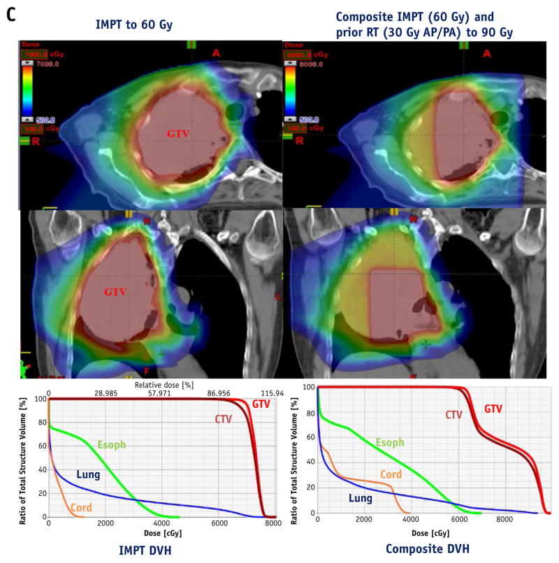
Purpose: Intensity modulated proton therapy (IMPT) can improve dose conformality and better spare normal tissue over passive scattering techniques, but range uncertainties complicate its use, particularly for moving targets. We report our early experience with IMPT for thoracic malignancies in terms of motion analysis and management, plan optimization and robustness, and quality assurance.

Methods and materials: Thirty-four consecutive patients with lung/mediastinal cancers received IMPT to a median 66 Gy(relative biological equivalence [RBE]). All patients were able to undergo definitive radiation therapy. IMPT was used when the treating physician judged that IMPT conferred a dosimetric advantage; all patients had minimal tumor motion (<5 mm) and underwent individualized tumor-motion dose-uncertainty analysis and 4-dimensional (4D) computed tomographic (CT)-based treatment simulation and motion analysis. Plan robustness was optimized by using a worst-case scenario method. All patients had 4D CT repeated simulation during treatment.

Results: IMPT produced lower mean lung dose (MLD), lung V5 and V20, heart V40, and esophageal V60 than did IMRT (P<.05) and lower MLD, lung V20, and esophageal V60 than did passive scattering proton therapy (PSPT) (P<.05). D5 to the gross tumor volume and clinical target volume was higher with IMPT than with intensity modulated radiation therapy or PSPT (P<.05). All cases were analyzed for beam-angle-specific motion, water-equivalent thickness, and robustness. Beam angles were chosen to minimize the effect of respiratory motion and avoid previously treated regions, and the maximum deviation from the nominal dose-volume histogram values was kept at <5% for the target dose and met the normal tissue constraints under a worst-case scenario. Patient-specific quality assurance measurements showed that a median 99% (range, 95% to 100%) of the pixels met the 3% dose/3 mm distance criteria for the γ index. Adaptive replanning was used for 9 patients (26.5%).

Conclusions: IMPT using 4D CT-based planning, motion management, and optimization was implemented successfully and met our quality assurance parameters for treating challenging thoracic cancers.


Conflict of interest statement

Conflict of interest: none.

Figures

Fig. 1
Fig. 1
Procedural flow chart for intensity modulated proton therapy (IMPT) quality assurance. 4D CT = 4-dimensional computed tomography; MFO = multifield optimization; SFO = single-field optimization.
Fig. 2
Fig. 2
(A) Comparison of dosimetric variables for intensity modulated proton therapy (IMPT), passive scattering proton therapy (PSPT), and intensity modulated radiation therapy (IMRT). Values shown are means of all patients. (B) Comparisons of dose-volume histogram (DVH), isodose distributions, and total mean lung dose for a representative patient who received chemotherapy with IMPT to 74 Gy(RBE) for stage III NSCLC. (C) Scans and DVHs for a patient with bulky stage III non–small cell lung cancer who had received urgent palliative photon therapy (30 Gy in 10 fractions using anterior/posterior fields, with spinal cord and esophagus receiving 30 Gy) followed by IMPT to 60 Gy in 30 fractions. The lesion could not be treated definitively with IMRT (because of a cumulative lung V5 >85%, not shown) or PSPT (significant amounts of esophagus would have received a cumulative dose of 80 to 90 Gy, not shown). (D) Scans and DVHs for a patient who had received photon therapy (60 Gy in 30 fractions) for small cell lung cancer, followed by IMPT to 45 Gy in 30 fractions twice a day with chemotherapy for an in-field recurrence. AP = anteroposterior; CTV = clinical target volume; GTV = gross tumor volume; MLD = mean lung dose; PA = posteroanterior; PET = positron emission tomography; RT = radiation therapy; Vx, volume of the cited organ (%) exposed to the indicated dose [in Gy(RBE)].
Fig. 2
Fig. 2
(A) Comparison of dosimetric variables for intensity modulated proton therapy (IMPT), passive scattering proton therapy (PSPT), and intensity modulated radiation therapy (IMRT). Values shown are means of all patients. (B) Comparisons of dose-volume histogram (DVH), isodose distributions, and total mean lung dose for a representative patient who received chemotherapy with IMPT to 74 Gy(RBE) for stage III NSCLC. (C) Scans and DVHs for a patient with bulky stage III non–small cell lung cancer who had received urgent palliative photon therapy (30 Gy in 10 fractions using anterior/posterior fields, with spinal cord and esophagus receiving 30 Gy) followed by IMPT to 60 Gy in 30 fractions. The lesion could not be treated definitively with IMRT (because of a cumulative lung V5 >85%, not shown) or PSPT (significant amounts of esophagus would have received a cumulative dose of 80 to 90 Gy, not shown). (D) Scans and DVHs for a patient who had received photon therapy (60 Gy in 30 fractions) for small cell lung cancer, followed by IMPT to 45 Gy in 30 fractions twice a day with chemotherapy for an in-field recurrence. AP = anteroposterior; CTV = clinical target volume; GTV = gross tumor volume; MLD = mean lung dose; PA = posteroanterior; PET = positron emission tomography; RT = radiation therapy; Vx, volume of the cited organ (%) exposed to the indicated dose [in Gy(RBE)].
Fig. 3
Fig. 3
Water-equivalent thickness (WET) analysis and beam angle selection. (A) Mean values of WET variation of the pixels in IGTV plotted as a function of beam angles. (B) WET analysis of a given beam angle of 160°, chosen based on minimal WET variation as shown in (A) along with tumor location for internal gross tumor volume (IGTV), with distal pixels calculated on reference (T0) and target (T50) phase scans; the difference between scans is shown as T50 minus T0. Histogram illustrates the percent volume differences in WET in the pixels within the IGTV was <5 mm (red vertical lines indicate S5mm). (C) Percentages of the voxels in IGTV with WET differences <5 mm (S5mm) are plotted as a function of beam angle. The chosen 160° beam angle was confirmed as being within the acceptability limit of ≥80%. A color version of this figure is available at www.redjournal.org.
Fig. 4
Fig. 4
Robustness analysis of an intensity modulated proton therapy plan. Solid lines indicate nominal dose-volume histogram calculated from the time-averaged computed tomographic scan; dotted lines indicate DVH calculated from the iso- or relative stopping power ratios shifted 9 scenarios. GTV = gross tumor volume; ITV = internal tumor volume.

References

    1. Chang JY, Cox JD. Improving radiation conformality in the treatment of non-small cell lung cancer. Semin Radiat Oncol. 2010;20:171–177. - PMC - PubMed
    1. Chang JY, Zhang X, Wang X, et al. Significant reduction of normal tissue dose by proton radiotherapy compared with three-dimensional conformal or intensity-modulated radiation therapy in stage I or stage III non-small-cell lung cancer. Int J Radiat Oncol Biol Phys. 2006;65:1087–1096. - PubMed
    1. Chang JY, Komaki R, Lu C, et al. Phase 2 study of high-dose proton therapy with concurrent chemotherapy for unresectable stage III nonsmall cell lung cancer. Cancer. 2011;117:4707–4713. - PMC - PubMed
    1. Koay EJ, Lege D, Mohan R, et al. Adaptive/nonadaptive proton radiation planning and outcomes in a phase II trial for locally advanced non-small cell lung cancer. Int J Radiat Oncol Biol Phys. 2012;84:1093–1100. - PMC - PubMed
    1. Gomez DR, Gillin M, Liao Z, et al. Phase 1 study of dose escalation in hypofractionated proton beam therapy for non-small cell lung cancer. Int J Radiat Oncol Biol Phys. 2013;86:665–670. - PMC - PubMed

Publication types

MeSH terms