

Actinobacillus pleuropneumoniae challenge in swine: diagnostic of lung alterations by infrared thermography
- PMID: 25260642
- PMCID: PMC4180138
- DOI: 10.1186/s12917-014-0199-2
Actinobacillus pleuropneumoniae challenge in swine: diagnostic of lung alterations by infrared thermography
Abstract
Background: Actinobacillus pleuropneumoniae (A.pp.) is the causative agent of porcine pleuropneumonia leading to high economic losses in the pig industry. Infrared thermography (IRT) of the thorax might offer a new method to select swine with lung alterations for further diagnostics. In this study 50 german landrace pigs were infected with A.pp. in an established model for respiratory tract disease, while 10 healthy pigs served as control animals. To avoid drift errors during IR measurements absolute skin temperatures and temperature differences between a thoracal and an abdominal region were assessed for its diagnostic validity.
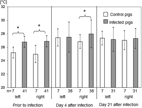
Results: IRT findings during the course of experimental A.pp.-infection were verified by computed tomography (CT) before and on days 4 and 21 after infection. Significant correlations were found between clinical scores, CT score and lung lesion score. Ambient temperature, body temperature and abdominal surface temperature were factors influencing the skin surface temperature of the thorax. On day 4 but not on day 21 after infection the right thoracal temperature was significantly higher and the difference between a thoracal region in the height of the left 10th vertebra and an abdominal region was significantly lower in infected pigs than in control pigs. At a cut off of 28°C of right thoracal temperature the specificity of the method was 100% (CI 95%: 69-100%) and the sensitivity 66% (CI 95%: 51-79%). At a cut off of 2°C temperature difference between thoracal and abdominal region on the left body site the specificity of the method was 100% (CI 95%: 69-100%) and the sensitivity 32% (CI 95%: 19-47%) with all control pigs detected negative. Orientation for lung biopsy by IRT resulted in 100% specificity and sensitivity (CI 95%: 69-100%) of bacteriological examination of tissue samples during the acute stage of infection.
Conclusion: IRT might be a valuable tool for the detection of inflammatory lung alterations in pigs, especially during the acute stage of infection and if ambient temperatures are constant during individual measurements. External and internal factors interfere with this method, so that its application in the field might be restricted to a selection of pigs for further diagnostic with adequate specificity.
Figures


References
-
- Hensel A, Ganter M, Kipper S, Krehon S, Wittenbrink MM, Petzoldt K. Prevalence of aerobic bacteria in bronchoalveolar lavage fluids from healthy pigs. Am J Vet Res. 1994;55:1697–1702. - PubMed
-
- Sorensen V, Jorsal SE, Mousing J. Diseases of the respiratory system. In: Straw BE, Zimmerman JJ, Allaire SD, Taylor DJ, editors. Diseases of Swine. 9. Washington: Blackwell Publishing; 2006. pp. 149–177.
-
- Brauer C, Hoeltig D, Hennig-Pauka I, Beyerbach M, Gasse H, Hewicker-Trautwein M, Gerlach GF, Waldmann KH. Computed tomography of the pig lung: an innovative approach to the definition of the pulmonary health status. Tierärztl Prax G. 2011;39:205–214. - PubMed
Publication types
MeSH terms
LinkOut - more resources
Full Text Sources
Other Literature Sources
Miscellaneous
