. 2014 Sep 27:14:252.
doi: 10.1186/s12870-014-0252-0.

Trans-Golgi network localized small GTPase RabA1d is involved in cell plate formation and oscillatory root hair growth

Trans-Golgi network localized small GTPase RabA1d is involved in cell plate formation and oscillatory root hair growth

Tobias Berson et al. BMC Plant Biol. .

Abstract

Background: Small Rab GTPases are important regulators of vesicular trafficking in plants. AtRabA1d, a member of the RabA1 subfamily of small GTPases, was previously found in the vesicle-rich apical dome of growing root hairs suggesting a role during tip growth; however, its specific intracellular localization and role in plants has not been well described.

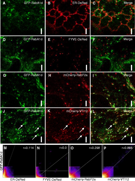
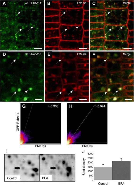
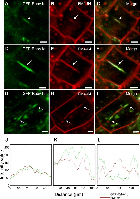
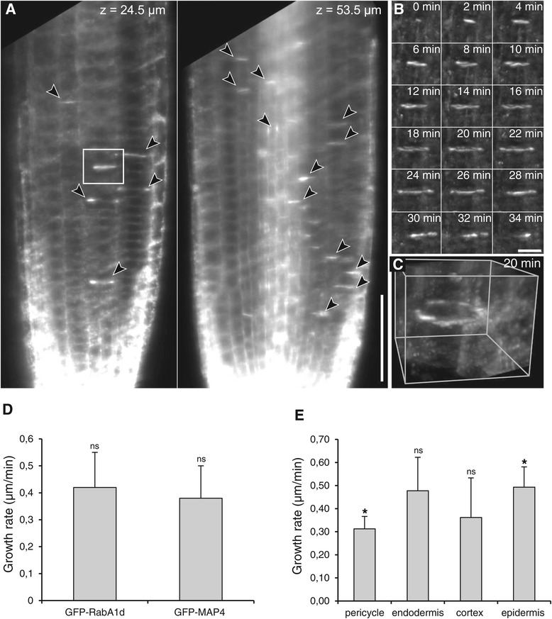
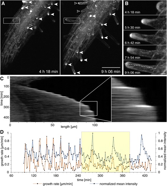
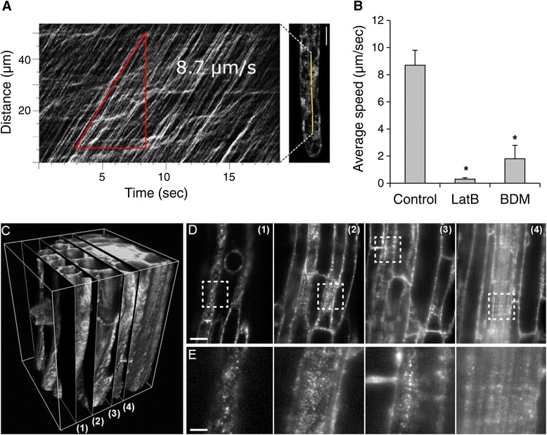
Results: The transient expression of 35S::GFP:RabA1d construct in Allium porrum and Nicotiana benthamiana revealed vesicular structures, which were further corroborated in stable transformed Arabidopsis thaliana plants. GFP-RabA1d colocalized with the trans-Golgi network marker mCherry-VTI12 and with early FM4-64-labeled endosomal compartments. Late endosomes and endoplasmic reticulum labeled with FYVE-DsRed and ER-DsRed, respectively, were devoid of GFP-RabA1d. The accumulation of GFP-RabA1d in the core of brefeldin A (BFA)-induced-compartments and the quantitative upregulation of RabA1d protein levels after BFA treatment confirmed the association of RabA1d with early endosomes/TGN and its role in vesicle trafficking. Light-sheet microscopy revealed involvement of RabA1d in root development. In root cells, GFP-RabA1d followed cell plate expansion consistently with cytokinesis-related vesicular trafficking and membrane recycling. GFP-RabA1d accumulated in disc-like structures of nascent cell plates, which progressively evolved to marginal ring-like structures of the growing cell plates. During root hair growth and development, GFP-RabA1d was enriched at root hair bulges and at the apical dome of vigorously elongating root hairs. Importantly, GFP-RabA1d signal intensity exhibited an oscillatory behavior in-phase with tip growth. Progressively, this tip localization dissapeared in mature root hairs suggesting a link between tip localization of RabA1d and root hair elongation. Our results support a RabA1d role in events that require vigorous membrane trafficking.

Conclusions: RabA1d is located in early endosomes/TGN and is involved in vesicle trafficking. RabA1d participates in both cell plate formation and root hair oscillatory tip growth. The specific GFP-RabA1d subcellular localization confirms a correlation between its specific spatio-temporal accumulation and local vesicle trafficking requirements during cell plate and root hair formation.


Figures

Figure 1
Figure 1
Subcellular localization of GFP-tagged RabA1d. Subcellular localization of GFP-RabA1d in cells of N. benthamiana. Co-vizualization with ER reporter ER-DsRed (A-C) showed partial association of GFP-RabA1d and cortical ER tubules. GFP-RabA1d colocalized with neither 2xFYVE-DsRed (D-F) nor with mCherry-RabF2a (G-I), markers for late endosomes/multi-vesicular bodies. Colocalization of GFP-RabA1d with mCherry-VTI12 representing a TGN marker (J-L). Intensity correlation scatterplots of GFP-Rab1Ad and ER-DsRed (M), FYVE-DsRed (N), mCherry-RabF2a (O), and mCherry-VTI12 (P). Pearson’s coefficient (r) was determined after Costes automatic threshold. Bars represent 3 μm in A-C and 5 μm in D-L.
Figure 2
Figure 2
GFP-RabA1d accumulates in BFA compartments and is upregulated by BFA treatment. Root cells of Arabidopsis stably transformed with 35S::GFP:RabA1d construct were analysed. GFP-RabA1d colocalized with early endocytotic compartments labeled by FM4-64 (A-C). After BFA treatment, both GFP-RabA1d and FM4-64 accumulated together in the core of BFA compartments (D-F). 2D-histogram intensity and correlation of GFP-Rab1Ad and FM4-64 early endocytotic compartments in root cells (G) and after BFA treatment (H). Pearson’s coefficient (r) was determined using Costes automatic threshold. BFA treatment induced RabA1d upregulation at protein level (I), upregulation of RabA1d was determined from comparison of 2-DE gels (arrow) and measured as increase of spot density (J). Bars represent 4 μm in A-C and 5 μm in D-F.
Figure 3
Figure 3
Localization of GFP-RabA1d during the cell plate formation in cytokinetic cells. During cell plate initiation, GFP-RabA1d localized in the mid plane of the dividing cell and completely colocalized with FM4-64 (A-C). In the early stage of growing cell plates, GFP-RabA1d still appeared in the mid-plane of the cell (D-F). During cytokinesis, GFP-RabA1d was accumulated mainly at the edges of growing cell plate (G-I). Intensity profile of GFP-RabA1d and FM4-64 signals during cell plate initiation and early stage of plate expansion (J,K), the two channels showed similar intensity and distribution; while in growing cell plate, GFP-RabA1d showed more intensity at the edges of growing cell plate than FM4-64 (L). Bars represent 2 μm.
Figure 4
Figure 4
Light-sheet live imaging of GFP-RabA1d accumulation during cell plate formation in the root meristem. Meristematic cells undergoing cytokinesis showed several locations of GFP-RabA1d accumulation observed in single sections of one time point at indicated depths inside the primary root (A), GFP-RabA1d accumulation in cell plates (arrowheads) of cytokinetic cells. The boxed area in A is enlarged shown in B. Detailed time-lapse imaging of GFP-RabA1d accumulation during single cell plate formation starting from spot like structures in the middle of the cell, followed by increased intensity during vesicle fusion and later formation of a ring-like structure at the margins in the growing cell plates (B). Three-dimensional reconstruction of the cell plate shown in (B) at time point 20 min (C). Comparison of cell plate growth rates in Arabidopsis plants stably transformed with in 35S::GFP:RabA1d and 35S::GFP:MAP4 constructs (ns: no significant difference) (D). Growth rates of cell plates in cells from different tissues (E). *significant difference from Wilcoxon rank sum test with Holm’s correction (p<0.05), ns: no significant difference. Bars represent 50 μm in A and 10 μm in B.
Figure 5
Figure 5
Light-sheet live imaging of GFP-RabA1d accumulation during root hair oscillatory tip growth. Maximum-intensity projections of two indicated time points (A). The actively growing root hairs showed a higher tip-accumulation (filled arrowheads) than the slowly expanding or mature root hairs (open arrowheads). The tip-accumulation of GFP-RabA1d increased in steady state actively growing root hairs (B, enlarged boxed area from A). The pixel line from B is plotted as a function of time (kymograph in C). The kymograph exhibited an oscillating behavior between lower and higher intensity (C). Fluorescence intensity measurements significantly correlated with the growth rates of the root hair (D). Bars represent 100 μm in A and 10 μm in B.
Figure 6
Figure 6
Motility of GFP-RabA1d vesicles in growing root hairs. Kymograph showing the motility of the GFP-RabA1d vesicles in root hairs (A). Estimation of the average speed of the GFP-RabA1d vesicles in root hairs treated with acto-myosin inhibitors, latrunculin B (LatB) and butane-dione-monoxime (BDM), respectively (B). Mobility and distribution of small RabA1d-positive TGN vesicles in diverse root tissues observed by light-sheet microscopy (C-E). 3D reconstruction of a stack of images (C). Single sections at different z-depths ((1): z = 0 μm, (2): z = 16.5 μm, (3): z = 36.8 μm, (4): z = 50 μm) are depicted in (D). The dashed boxed area is enlarged in (E). Bars represent 20 μm in D and 5 μm in E.

References

    1. Campanoni P, Blatt MR. Membrane trafficking and polar growth in root hairs and pollen tubes. J Exp Bot. 2007;58:65–74. doi: 10.1093/jxb/erl059. - DOI - PubMed
    1. Cole RA, Fowler JE. Polarized growth: maintaining focus on the tip. Curr Opin Plant Biol. 2006;9:579–588. doi: 10.1016/j.pbi.2006.09.014. - DOI - PubMed
    1. Zárský V, Cvrčková F, Potocký M, Hála M. Exocytosis and cell polarity in plants - exocyst and recycling domains. New Phytol. 2009;183:255–272. doi: 10.1111/j.1469-8137.2009.02880.x. - DOI - PubMed
    1. Nielsen E, Cheung AY, Ueda T. The regulatory RAB and ARF GTPases for vesicular trafficking. Plant Physiol. 2008;147:1516–1526. doi: 10.1104/pp.108.121798. - DOI - PMC - PubMed
    1. Saito C, Ueda T. Functions of RAB and SNARE proteins in plant life. Int Rev Cell Mol Biol. 2009;274:183–233. doi: 10.1016/S1937-6448(08)02004-2. - DOI - PubMed

Publication types

MeSH terms

Substances

LinkOut - more resources