

Reverse genetics system for studying human rhinovirus infections
- PMID: 25261313
- PMCID: PMC4422385
- DOI: 10.1007/978-1-4939-1571-2_12
Reverse genetics system for studying human rhinovirus infections
Abstract
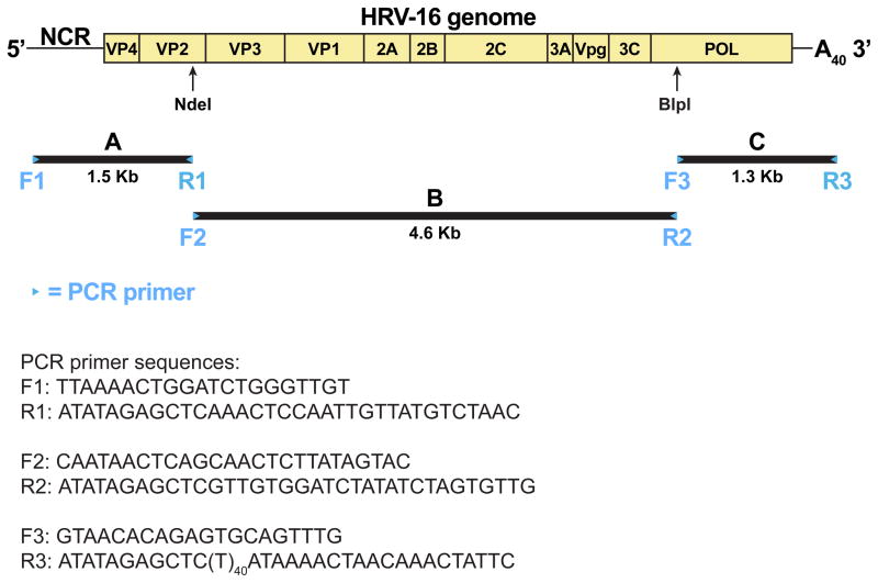
Human rhinovirus (HRV) contains a 7.2 kb messenger-sense RNA genome which is the template for reproducing progeny viruses after it enters the cytoplasm of a host cell. Reverse genetics refers to the regeneration of progeny viruses from an artificial cDNA copy of the RNA genome of an RNA virus. It has been a powerful molecular genetic tool for studying HRV and other RNA viruses because the artificial DNA stage makes it practical to introduce specific mutations into the viral RNA genome. This chapter uses HRV-16 as the model virus to illustrate the strategy and methods for constructing and cloning the artificial cDNA copy of a full-length HRV genome, identifying the infectious cDNA clone isolates, and selecting the most vigorous cDNA clone isolate to serve as the standard parental clone for future molecular genetic study of the virus.
Figures
References
-
- Turner RB, Lee W-M. In: Clinical Virology. Richman DD, Whitley RJ, Hayden FG, editors. ASM Press; Washington, D.C: 2009. pp. 1063–82.
-
- Rueckert R. In: Fields Virology. 3. Fields BN, Knipe DM, Howley PM, et al., editors. Lippincott-Raven Publishers; Philadelphia: 1996. pp. 609–54.
-
- Taniguchi T, Palmieri M, Weissmann C. Nature. 1978;274:223–8. - PubMed
MeSH terms
Substances
Grants and funding
LinkOut - more resources
Full Text Sources
Other Literature Sources
