

Unrecognized preclinical Alzheimer disease confounds rs-fcMRI studies of normal aging
- PMID: 25261500
- PMCID: PMC4223085
- DOI: 10.1212/WNL.0000000000000939
Unrecognized preclinical Alzheimer disease confounds rs-fcMRI studies of normal aging
Abstract
Objective: To determine whether, and to what degree, preclinical Alzheimer disease (AD) confounds studies of healthy aging where "healthy" is based on cognitive normality alone.
Methods: We examined the effects of preclinical AD in cognitively normal older individuals using resting-state functional connectivity MRI. We investigated 2 groups of cognitively normal participants: one group with evidence of preclinical AD as assessed by CSF markers of AD and the other group with normal CSF biomarkers.
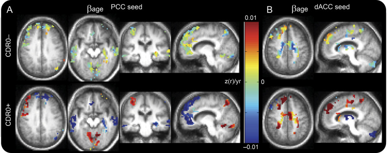
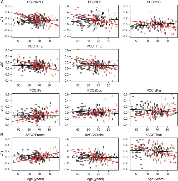
Results: There were significant interactions between age and biomarker status in the default-mode, dorsal attention, and salience resting-state networks. In the group with evidence of preclinical AD, there were dramatic changes in functional connectivity with age. In the group without evidence of preclinical AD, those changes were greatly attenuated. In most regions with significant interactions of age and biomarker status, the age-related change in functional connectivity in the normal biomarker group was indistinguishable from zero.
Conclusions: These results suggest that preclinical AD accounts for a substantial portion of the reported effects of aging in the extant functional connectivity literature.
© 2014 American Academy of Neurology.
Conflict of interest statement
M. Brier, J. Thomas, A. Snyder, and L. Wang report no disclosures relevant to the manuscript. A. Fagan consults for Roche and Lilly USA. T. Benzinger has a research grant from Avid Radiopharmaceuticals (a wholly owned subsidiary of Eli Lilly), and has received other support from the National Multiple Sclerosis Society (travel for grant review). J. Morris has participated in drug trials with Janssen Immunotherapy, Pfizer, and Eli Lilly, and has received honoraria from the Charles A. Dana Foundation, Eli Lilly, and Eisai. B. Ances reports no disclosures relevant to the manuscript. Go to
Figures
Comment in
-
Alzheimer disease: Altered functional connectivity in preclinical dementia.Nat Rev Neurol. 2014 Nov;10(11):609. doi: 10.1038/nrneurol.2014.195. Epub 2014 Oct 21. Nat Rev Neurol. 2014. PMID: 25330722 No abstract available.
References
-
- Koch W, Teipel S, Mueller S, et al. . Effects of aging on default mode network activity in resting state fMRI: does the method of analysis matter? Neuroimage 2010;51:280–287. - PubMed
-
- Knopman DS, Parisi JE, Salviati A, et al. . Neuropathology of cognitively normal elderly. J Neuropathol Exp Neurol 2003;62:1087–1095. - PubMed
Publication types
MeSH terms
Substances
Grants and funding
- P01AG026276/AG/NIA NIH HHS/United States
- P01 AG026276/AG/NIA NIH HHS/United States
- R01NRO14449/PHS HHS/United States
- P01AG03991/AG/NIA NIH HHS/United States
- R01NR012907/NR/NINR NIH HHS/United States
- R01 NR012907/NR/NINR NIH HHS/United States
- NSO6833/PHS HHS/United States
- UL1 TR000448/TR/NCATS NIH HHS/United States
- U19AG032438/AG/NIA NIH HHS/United States
- P30NSO48056/PHS HHS/United States
- R01NRO12657/PHS HHS/United States
- PSOAG05681/PHS HHS/United States
- P01 AG003991/AG/NIA NIH HHS/United States
- P50 AG005681/AG/NIA NIH HHS/United States
LinkOut - more resources
Full Text Sources
Other Literature Sources
Medical