. 2014 Dec;80(24):7551-60.
doi: 10.1128/AEM.02676-14. Epub 2014 Sep 26.

Dynamics of gut microbiota in autoimmune lupus

Affiliations

Dynamics of gut microbiota in autoimmune lupus

Husen Zhang et al. Appl Environ Microbiol. 2014 Dec.

Abstract

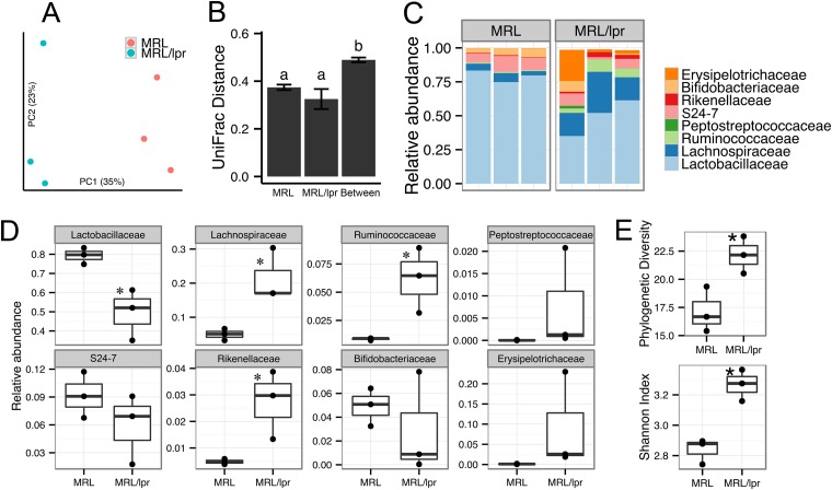
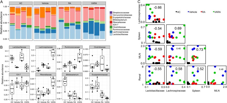
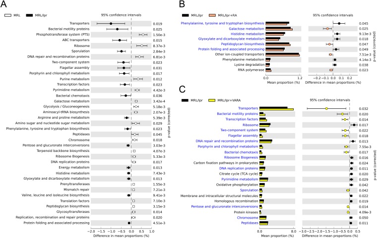
Gut microbiota has been recognized as an important environmental factor in health, as well as in metabolic and immunological diseases, in which perturbation of the host gut microbiota is often observed in the diseased state. However, little is known on the role of gut microbiota in systemic lupus erythematosus. We investigated the effects of host genetics, sex, age, and dietary intervention on the gut microbiome in a murine lupus model. In young, female lupus-prone mice resembling women at childbearing age, a population with the highest risk for lupus, we found marked depletion of lactobacilli, and increases in Lachnospiraceae and overall diversity compared to age-matched healthy controls. The predicted metagenomic profile in lupus-prone mice showed a significant enrichment of bacterial motility- and sporulation-related pathways. Retinoic acid as a dietary intervention restored lactobacilli that were downregulated in lupus-prone mice, and this correlated with improved symptoms. The predicted metagenomes also showed that retinoic acid reversed many lupus-associated changes in microbial functions that deviated from the control. In addition, gut microbiota of lupus-prone mice were different between sexes, and an overrepresentation of Lachnospiraceae in females was associated with an earlier onset of and/or more severe lupus symptoms. Clostridiaceae and Lachnospiraceae, both harboring butyrate-producing genera, were more abundant in the gut of lupus-prone mice at specific time points during lupus progression. Together, our results demonstrate the dynamics of gut microbiota in murine lupus and provide evidence to suggest the use of probiotic lactobacilli and retinoic acid as dietary supplements to relieve inflammatory flares in lupus patients.


Figures

FIG 1
FIG 1
Differences in gut microbiota between healthy and lupus-prone mice. (A) Microbiota separation on the first two principal coordinates calculated from unweighted UniFrac distances. MRL, healthy control mice; MRL/lpr, lupus-prone mice. Fecal samples from 5-week-old female mice were used. The number of sequences in each of the six samples was 7,800. (B) UniFrac distances within and between mouse strains. Error bars indicate the standard errors of the mean for n = 3 (within MRL), n = 3 (within MRL/lpr), and n = 9 (between the two strains). Statistical significance was determined by one-way ANOVA, followed by pairwise t test with TukeyHSD-adjusted P values. (C) Taxonomy breakdown at the family level for abundant (>0.1%) bacterial OTU. (D) Bacterial abundance comparison between MRL and MRL/lpr microbiota. (E) Diversity indices. Statistical comparison was based on an unpaired Student t test (n = 3 in each group; *, P < 0.05; **, P < 0.01).
FIG 2
FIG 2
Microbiota dynamics associated with sex and age. (A) Sex-associated microbiota differences. Colonic contents from 14-week-old MRL and MRL/lpr mice were used. Statistical significance was determined by two-way ANOVA, followed by pairwise t test with TukeyHSD-adjusted significance (n = 4 in each group). (B and C) Age-associated microbiota differences. (B) Fecal microbiota separation on the first two principal coordinates calculated from unweighted UniFrac distances. b6, female healthy control mice; b6/lpr, female lupus-prone mice. (C) Time-dependent changes of fecal microbiota in b6 and b6/lpr strains. Abundant bacterial OTU (>0.1%) were summarized (n = 3 per group).
FIG 3
FIG 3
Changes of bacterial abundance with vitamin A treatments. (A) Taxonomy breakdown at the family level abundant (>0.1%) bacterial OTU. HC, MRL healthy control treated with canola oil; vehicle, lupus-prone MRL/lpr treated with canola oil; RA, MRL/lpr treated with retinoic acid (6 mg/kg of body weight); VARA, MRL/lpr treated with retinol (6 mg retinol/kg of body weight) and retinoic acid (0.6 mg/kg of body weight, which facilitates retinol storage). All mice were female and treated orally from 6 to 14 weeks of age. Mice were sacrificed at 14 weeks of age and colonic contents were collected (n = 4 per group). (B) Comparison of individual families shown in panel A. Statistical significance was determined by one-way ANOVA, followed by a pairwise t test with TukeyHSD-adjusted significance. (C) Correlation between bacterial abundance and lupus disease indices. Spleen, spleen weight in grams; MLN, mesenteric lymph node weight in grams; renal, renal function estimated from proteinuria and glomerular scores as described in Materials and Methods. Correlation coefficients are shown on each individual plot. For example, the correlation between Lactobacillaceae and Lachnospiraceae was −0.66. Spearman's correlation tests were used for pairs involving ranked renal function data; all other correlations were calculated by using the Pearson method. Raw data can be found in the supplemental material.
FIG 4
FIG 4
Bacterial metagenomes predicted from microbial community identities. Gene functional categories were from level 3 of KEGG pathways. Gene functions with a significant difference are shown (P < 0.05). (A) Differences between MRL control and lupus-prone MRL/lpr mice. The effect size was 0.10%. ABC transporters, ATP-binding cassette transport systems. (B) Effect of RA treatment on lupus-prone bacterial metagenomes. The effect size was 0.02%. (C) Effect of retinol treatment (VARA) on lupus-prone bacterial metagenomes. The effect size was 0.08%.

References

    1. Ley RE, Peterson DA, Gordon JI. 2006. Ecological and evolutionary forces shaping microbial diversity in the human intestine. Cell 124:837–848. 10.1016/j.cell.2006.02.017. - DOI - PubMed
    1. Kamada N, Seo SU, Chen GY, Nunez G. 2013. Role of the gut microbiota in immunity and inflammatory disease. Nat. Rev. Immunol. 13:321–335. 10.1038/nri3430. - DOI - PubMed
    1. Kuhn KA, Stappenbeck TS. 2013. Peripheral education of the immune system by the colonic microbiota. Semin. Immunol. 25:364–369. 10.1016/j.smim.2013.10.002. - DOI - PMC - PubMed
    1. Strober W. 2013. Impact of the gut microbiome on mucosal inflammation. Trends Immunol. 34:423–430. 10.1016/j.it.2013.07.001. - DOI - PMC - PubMed
    1. Yeoh N, Burton JP, Suppiah P, Reid G, Stebbings S. 2013. The role of the microbiome in rheumatic diseases. Curr. Rheumatol. Rep. 15:314. 10.1007/s11926-012-0314-y. - DOI - PubMed

Publication types