Review
. 2015 Apr:322:77-88.
doi: 10.1016/j.heares.2014.09.009. Epub 2014 Sep 28.

Importance of cochlear health for implant function

Affiliations
Review

Importance of cochlear health for implant function

Bryan E Pfingst et al. Hear Res. 2015 Apr.

Abstract

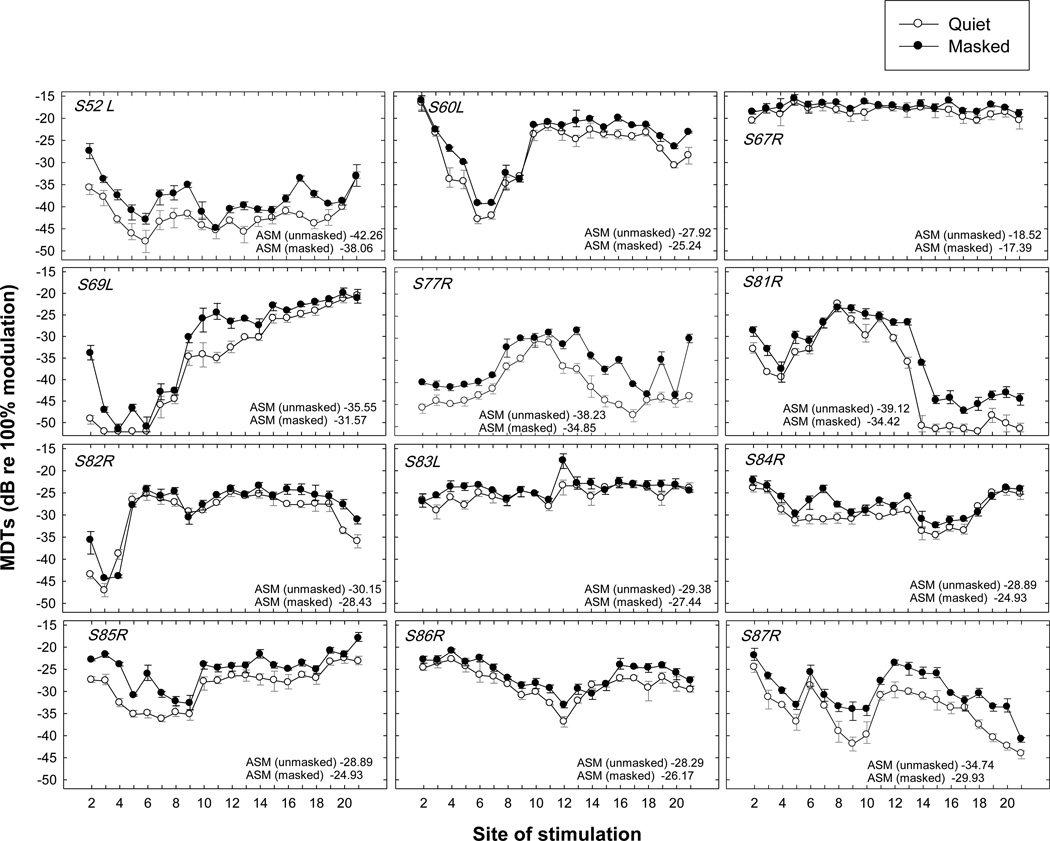
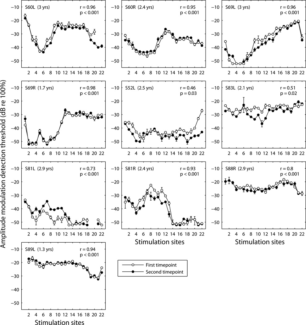
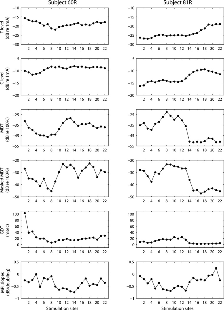
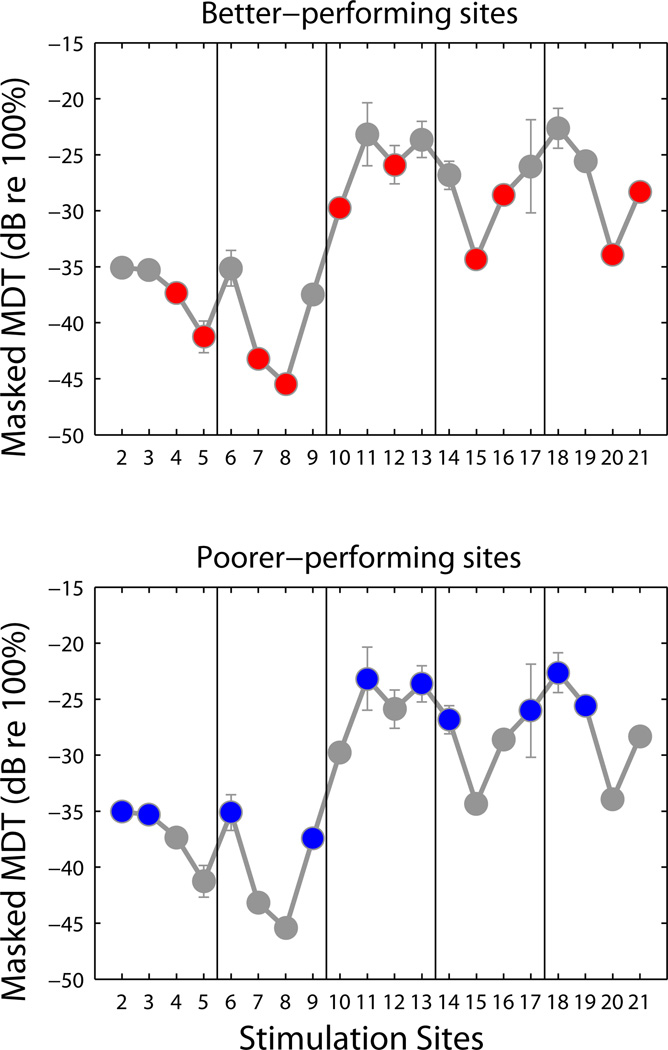
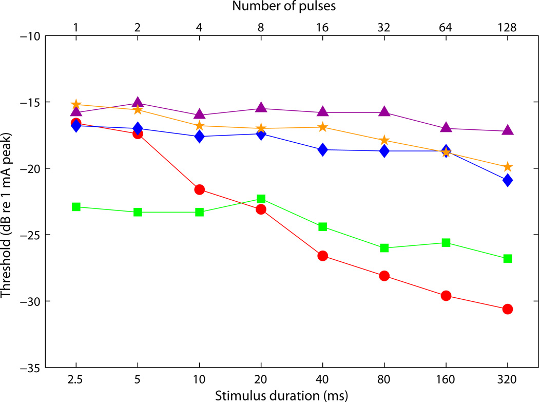
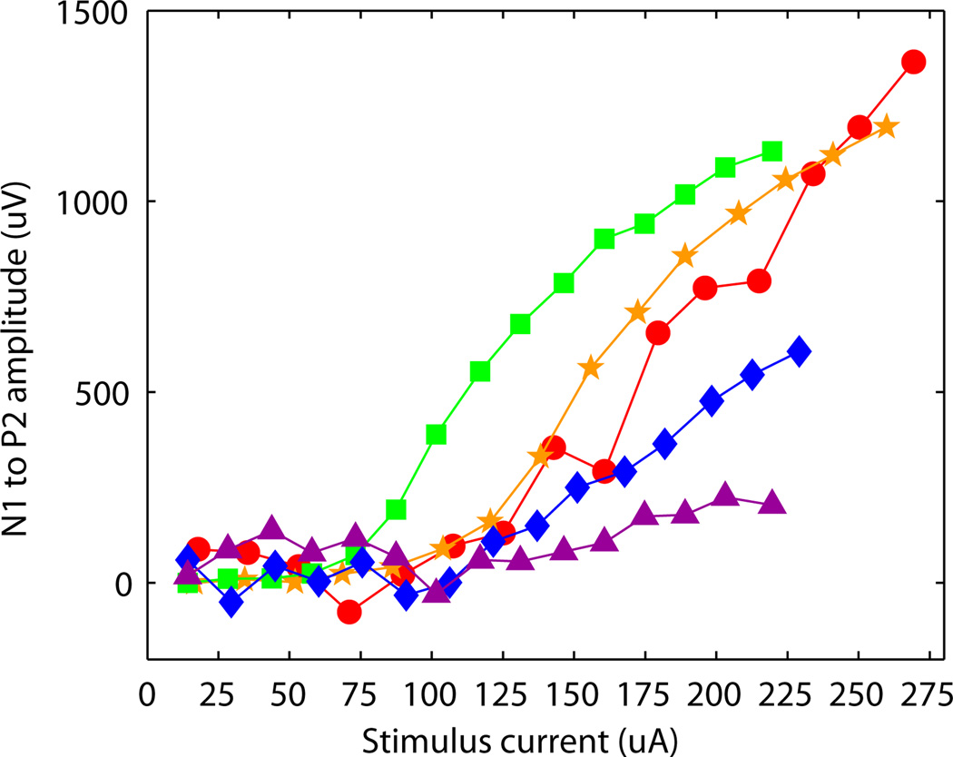
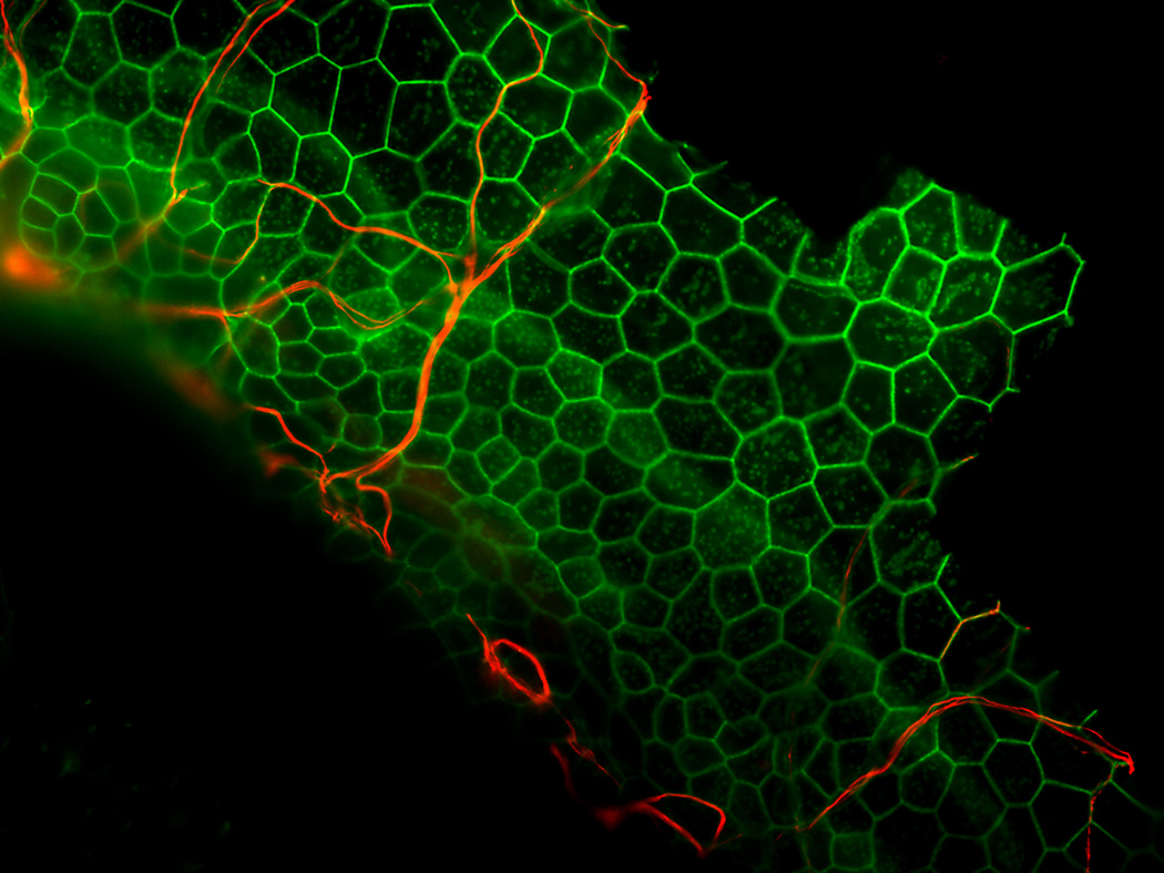
Amazing progress has been made in providing useful hearing to hearing-impaired individuals using cochlear implants, but challenges remain. One such challenge is understanding the effects of partial degeneration of the auditory nerve, the target of cochlear implant stimulation. Here we review studies from our human and animal laboratories aimed at characterizing the health of the implanted cochlea and the auditory nerve. We use the data on cochlear and neural health to guide rehabilitation strategies. The data also motivate the development of tissue-engineering procedures to preserve or build a healthy cochlea and improve performance obtained by cochlear implant recipients or eventually replace the need for a cochlear implant. This article is part of a Special Issue entitled <Lasker Award>.


Figures

Figure 1
Figure 1
Modulation detection thresholds (MDTs) as a function of stimulation site for 12 human subjects with 22-electrode cochlear implants. Stimulation sites (electrodes) are numbered 1 to 22 from the basal end to the apical end of the cochlear implant electrode array. MDTs represent the minimum modulation depth at which a subject can discriminate an amplitude-modulated pulse train from a non-modulated pulse train. Functions for MDTs in quiet (open circles) and in the presence of non-modulated masker on an adjacent, more apical, electrode (filled circles) are shown. Larger negative values indicate better performance. Across-site mean (ASM) MDTs are shown in the lower right corner of each panel. Error bars show ranges of values from three estimates at each site. Data are from Garadat et al., 2012.
Figure 2
Figure 2
Stability of modulation detection thresholds over time. Data for 10 ears are shown with the subject and ear designation given in the upper left corner of each panel. Modulation detection thresholds were measured at all available stimulation sites at two timepoints with the time elapsed between the two timepoints (in years) shown in each panel. The first and second sets of data are shown in different symbols: open symbols for the first timepoint and filled symbols for the second timepoint. Each data point represents the mean of two measurements at a given timepoint with error bars representing the range of the data. Statistics for correlation across the electrode array between the data at the first and second timepoints are shown in the upper–right corner of each panel. For these correlations the means of the two measurements at each site at the first timepoint were correlated with the means of the two measurements at each site at the second timepoint.
Figure 3
Figure 3
Across-site patterns for six psychophysical measures in two subjects (one subject per column). The measures are from the top absolute detection threshold (T level), maximal comfortable level (C level), modulation detection threshold (MDT), modulation detection threshold in presence of a masker on the electrode immediately basal to the probe (masked MDT), gap-detection threshold (GDT), and slope of the multipulse integration function (MPI slopes). All measures were obtained using an MP 1+2 configuration. The supra-threshold functions were measured at levels corresponding to 50% of the site’s dynamic range. More information about methods used to collect these types of data can be obtained from previous publications from this laboratory. For T levels, C levels and MDTs and masked MDTs, see Garadat et al., 2012 or Zhou and Pfingst, 2012. For GDTs see Garadat and Pfingst, 2011. For MPI slopes see Zhou et al., 2012 or 2014a).
Figure 4
Figure 4
Examples of a site-selection strategy to test the relevance of MDTs for speech recognition, based on the study by Garadat and colleagues (2012). Two 10-electrode processor maps were created: one with 10 of the better-performing sites (red circles in the top panel) and one with 10 poorer-performing sites (blue circles in the bottom panel). To maintain coverage of the full range of place-pitch information, the electrode array (electrodes 2 through 21) was divided into 5 segments and two sites were selected from each segment. Speech recognition, particularly sentence recognition in noise, was better in most subjects when the map with the better-performing sites was used.
Figure 5
Figure 5
Examples of temporal integration (TI) functions (psychophysical detection thresholds as a function of pulse-train duration) from five animals with various levels of spiral ganglion neuron (SGN) preservation near the implant as detailed in Table 1.
Figure 6
Figure 6
Examples of ECAP amplitude-growth functions from the same five animals for which TI data are shown in Figure 5. The N1 to P2 ECAP amplitude (µV) was used because P1 was usually obscured by stimulus artifact. Growth function slopes, but not thresholds were correlated with the degree of SGN survival as detailed in Table 1.
Figure 7
Figure 7
Example of neurite growth toward the basilar membrane area in a deaf, neurotrophin treated ear. A whole-mount of the basal turn of the guinea pig cochlea stained for neurofilaments (red) and actin (green) and viewed with epi-fluorescence is shown. The ear was deafened with neomycin, injected with AAV. NTF-3 a week later and obtained for histology 3 months after that. The auditory epithelium does not contain differentiated hair cells or supporting cells. Instead, it is composed of flat or cuboidal simple epithelium. Nerve fibers are seen entering the epithelium and traversing the epithelial cells. This experiment was similar to that reported by Shibata and colleagues (2010) except that AAV was used in this case instead of Ad as the vector for gene therapy.

Similar articles

Cited by

References

    1. Bas E, Van De Water TR, Lumbreras V, Rajguru S, Goss G, Hare JM, Goldstein BJ. Adult human nasal mesenchymal-like stem cells restore cochlear spiral ganglion neurons after experimental lesion. Stem Cells Devel. 2014;23:502–514. - PMC - PubMed
    1. Bierer JA. Threshold and channel interaction in cochlear implant users: Evaluation of the tripolar electrode configuration. J. Acoust. Soc. Am. 2007;121:1642–1653. - PubMed
    1. Bierer JA, Faulkner KF. Identifying cochlear implant channels with poor electrode-neuron interface: Partial tripolar, single-channel thresholds and psychophysical tuning curves. Ear Hear. 2010;31:247–258. - PMC - PubMed
    1. Budenz CL, Pfingst BE, Raphael Y. The use of neurotrophin therapy in the inner ear to augment cochlear implantation outcomes. Anat. Rec. 2012;295:1896–1908. - PMC - PubMed
    1. Chen W, Jongkamonwiwat N, Abbas L, Eshtan SJ, Johnson SL, Kuhn S, Milo M, Thurlow JK, Andrews PW, Marcotti W, Moore HD, Rivolta MN. Restoration of auditory evoked responses by human ES-cell-derived otic progenitors. Nature. 2012;490:278–282. - PMC - PubMed

Publication types