. 2014 Sep 28:9:39.
doi: 10.1186/1750-1326-9-39.

Histone deacetylase 3 (HDAC3) plays an important role in retinal ganglion cell death after acute optic nerve injury

Affiliations

Histone deacetylase 3 (HDAC3) plays an important role in retinal ganglion cell death after acute optic nerve injury

Heather M Schmitt et al. Mol Neurodegener. .

Abstract

Background: Optic nerve damage initiates a series of early atrophic events in retinal ganglion cells (RGCs) that precede the BAX-dependent committed step of the intrinsic apoptotic program. Nuclear atrophy, including global histone deacetylation, heterochromatin formation, shrinkage and collapse of nuclear structure, and the silencing of normal gene expression, comprise an important obstacle to overcome in therapeutic approaches to preserve neuronal function. Several studies have implicated histone deacetylases (HDACs) in the early stages of neuronal cell death, including RGCs. Importantly, these neurons exhibit nuclear translocation of HDAC3 shortly after optic nerve damage. Additionally, HDAC3 activity has been reported to be selectively toxic to neurons.

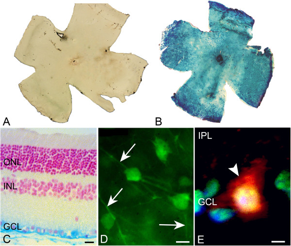
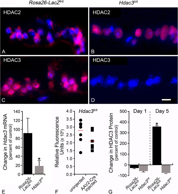
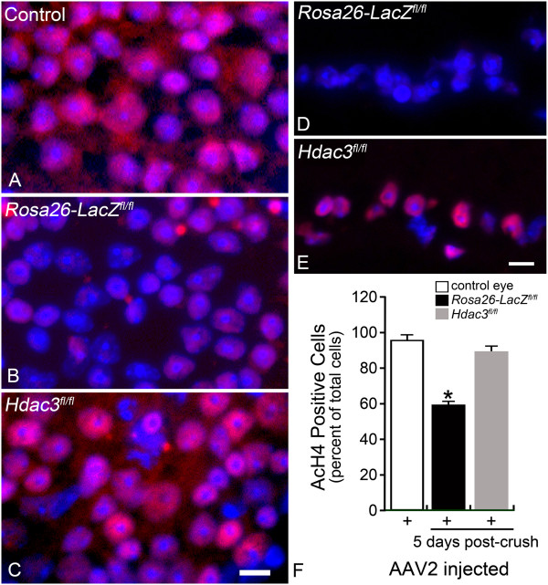
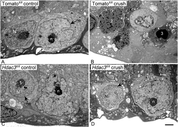
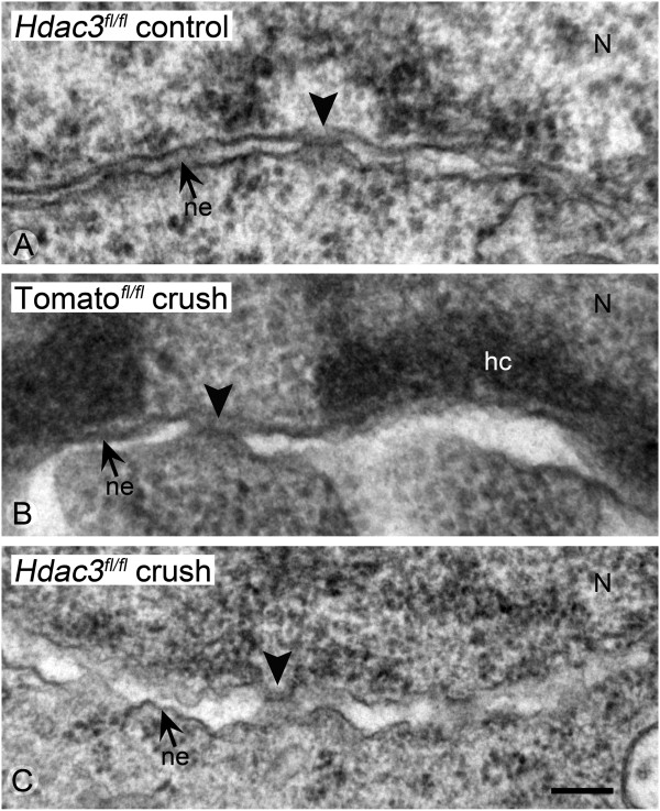
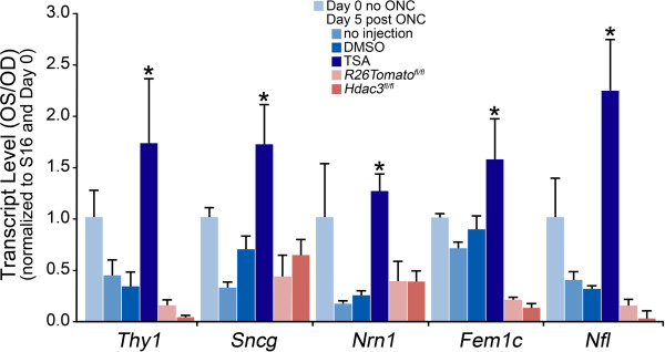
Results: RGC-specific conditional knockout of Hdac3 was achieved by transducing the RGCs of Hdac3fl/fl mice with an adeno-associated virus serotype 2 carrying CRE recombinase and GFP (AAV2-Cre/GFP). Controls included similar viral transduction of Rosa26fl/fl reporter mice. Optic nerve crush (ONC) was then performed on eyes. The ablation of Hdac3 in RGCs resulted in significant amelioration of characteristics of ONC-induced nuclear atrophy such as H4 deacetylation, heterochromatin formation, and the loss of nuclear structure. RGC death was also significantly reduced. Interestingly, loss of Hdac3 expression did not lead to protection against RGC-specific gene silencing after ONC, although this effect was achieved using the broad spectrum inhibitor, Trichostatin A.

Conclusion: Although other HDACs may be responsible for gene expression changes in RGCs, our results indicate a critical role for HDAC3 in nuclear atrophy in RGC apoptosis following axonal injury. This study provides a framework for studying the roles of other prevalent retinal HDACs in neuronal death as a result of axonal injury.


Figures

Figure 1
Figure 1
AAV2-Cre/GFP transduces RGCs in Rosa26-LacZ fl/fl and Rosa26-Tomato fl/fl mice. (A-B) Retinal whole mounts from non-injected (A) and injected (B) eyes of Rosa26-LacZ fl/fl mice were taken 8 weeks following injection. X-Gal staining indicates global reporter gene expression in injected eyes. (C) Retinal section taken from an injected eye of Rosa26-LacZ fl/fl illustrates that X-Gal staining is restricted to cells of the ganglion cell layer (GCL) (Scale bar: 20 μm) and is not present in the outer nuclear layer (ONL) and inner nuclear layer (INL). (D) GFP fluorescence carried by AAV2-Cre/GFP is found in the RGC somas and axons (indicated by arrows) in a retinal whole mount (Scale bar: 10 μm). (E) AAV2-Cre/GFP tropism to RGCs in the GCL, and not to the inner plexiform layer (IPL) is shown by nuclear BRN3A (green), TOMATO (red), and DAPI (blue) co-labeling in a retinal section (arrowhead). (Scale bar: 4 μm).
Figure 2
Figure 2
Intravitreal AAV2-Cre/GFP injection achieved conditional knockout of the Hdac3 gene. (A-B) TEXAS RED labeling of HDAC2 in Rosa26-LacZ fl/fl and Hdac3 fl/fl AAV2-Cre/GFP injected eyes at 5 days post ONC illustrates nuclear localization of this HDAC in cells of the GCL of both retinas. (C-D) TEXAS RED labeling of HDAC3 in Rosa26-LacZ fl/fl and Hdac3 fl/fl AAV2-Cre/GFP injected eyes at 5 days post ONC demonstrates the lack of HDAC3 in the Hdac3 fl/fl RGCs (Scale bar: 15 μm). (E) QPCR was used to determine abundance of Hdac3 mRNA from pooled retinas of 5 Rosa26-LacZ fl/fl and Hdac3 fl/fl mice at 4 weeks following AAV2-Cre/GFP injection. In uncrushed retinas, the abundance of Hdac3 mRNA was significantly lower in Hdac3 cKO retinas compared to Rosa26-LacZ fl/fl retinas (*P ≤ 0.05). (F) Protein from individual retinas of AAV2-Cre/GFP injected (4-week incubation) and uninjected Hdac3 fl/fl mice showed an overall decrease in protein levels in Hdac3 cKO mice, although there was no significant difference in mean abundance (P > 0.05). (G) Pooled retinal protein from 5 AAV2-Cre/GFP injected Rosa26-LacZ fl/fl and Hdac3 fl/fl mice at 1 and 5 days post ONC were run on a Western blot. ONC results in an increase in HDAC3 accumulation in Rosa26-LacZ fl/fl mice by 5 days, but this response is significantly abrogated in Hdac3 fl/fl animals (*P ≤ 0.05) when comparing Rosa26-LacZ fl/fl and Hdac3 fl/fl mice at 5 days.
Figure 3
Figure 3
Hdac3 cKO ameliorated global deacetylation following RGC injury. (A-C) Retinal whole mounts were stained for AcH4 (red) in Rosa26-LacZ fl/fl and Hdac3 fl/fl AAV2-Cre/GFP injected and uninjected eyes at 5 days following optic nerve crush. The control retina was an uninjected and uncrushed Hdac3 fl/fl retina, which exhibited widespread AcH4 labeling (Scale bar: 10 μm). Nuclei absent of AcH4 staining were present only in the Rosa26-LacZ fl/fl retina after ONC. (D-E) Fluorescent microscopy of the retinal sections showed that the Hdac3 cKO GCL retained visibly more AcH4 labeled cells than the Rosa26-LacZ fl/fl GCL (Scale bar: 15 μm). (F) Cell counts in the GCL indicated Hdac3 cKO retinas at 5 days post ONC, retained AcH4 levels comparable to control retinas (P > 0.05), while Rosa26-LacZ fl/fl retinas exhibited about a 40% decrease in AcH4 labeled cells in the GCL compared to control retinas (*P ≤ 0.05).
Figure 4
Figure 4
Rosa26-Tomato fl/fl retinas exhibited significantly more pyknotic nuclei at 5 days following ONC when compared to Hdac3 fl/fl cKO and control retinas. (A) A section of the retina from a Rosa26-Tomato fl/fl mouse, 5 days after ONC, to show an exemplar of the heterochromatin scoring system. (B) Scoring data from masked observers is depicted by a bar graph. Significantly more heterochromatic and pyknotic nuclei were detected in Rosa26-Tomato fl/fl control crushed retinas when compared to uncrushed and Hdac3 cKO crushed retinas (*P < 0.05). Heterochromatin score of 1 = healthy cell with euchromatic nucleus and well-formed nucleolus, 2 = cell with partial apoptotic heterochromatin formation, and 3 = cell with completely heterochromatic (pyknotic) fragmented nuclei. (Scale bar: 10 μm).
Figure 5
Figure 5
Hdac3 cKO prevented apoptotic heterochromatin formation in TUJ-1 positive cells in the GCL 5 days post ONC. (A-C) The GCL of whole mounts from a Rosa26-LacZ fl/fl control eye, a Rosa26-LacZ fl/fl crushed eye, and an Hdac3 fl/fl crushed eye stained with the TUJ-1 monoclonal antibody. Nuclear morphology was stained using DAPI. The DAPI staining of a representative cells is shown in the inset in the lower right corner of each panel. Nuclei of TUJ-1 positive cells in control (A) and Hdac3 fl/fl crushed retinas (B) typically exhibited a normal appearance with minimal heterochromatin and a prominent nucleolus. (C) In Rosa26-LacZ fl/fl retinas after ONC, however, some nuclei clearly showed condensed staining and were fragmented. (D) Graphical representation of cell counts indicating a significantly higher percentage of TUJ-1 positive apoptotic cells in Rosa26-LacZ fl/fl crushed eyes when compared to crushed Hdac3 cKO and control eyes (*P ≤ 0.001). (Scale bars: 10 μm).
Figure 6
Figure 6
Hdac3 cKO blocked heterochromatin formation in the GCL 5 days following ONC. (A-B) TEM images of cells in the GCL of a Rosa26-Tomato fl/fl control eye and AAV2-Cre/GFP injected eye, 5 days after ONC. Prominent heterochromatin formation is evident in the GCL of Rosa26-Tomato fl/fl crushed retinas. (C-D) TEM images of cells in the GCL of an Hdac3 fl/fl control eye and Hdac3 cKO crushed eye indicate no heterochromatin formation in the GCL as a result of ONC. Nuclei with characteristics of the morphological scores described in Figure 4 are indicated. N = nucleus, n = nucleolus, ne = nuclear envelope, and m = Müller endfoot. (Scale bar: 3 μm).
Figure 7
Figure 7
Hdac3 cKO in the RGCs prevented the breakdown of the nuclear envelope and pore integrity following ONC. (A-C) TEM images were taken of nuclear envelopes from Hdac3 fl/fl control retinas, Rosa26-Tomato fl/fl crushed retinas, and Hdac3 fl/fl crushed retinas. The nuclear lamina is situated at the top of each micrograph. (A) Example of the well-defined inner and outer membranes of the nuclear envelope and normal pore structure of a healthy cell. (B) Example of the nuclear envelope from a cell in a Rosa26-Tomato fl/fl retina 5 days after ONC. The nucleus exhibits formation of electron-dense heterochromatin localized to the inner surface of the nuclear envelope, and the intermembrane space has expanded. (C) The nuclear envelope of a presumptive RGC in an Hdac3 fl/fl cKO mouse after ONC. The chromatin appears euchromatic and the nuclear envelop exhibits pore structures, although the apposition of the inner and outer membranes appears wavy. Arrowheads = nuclear pores, small arrows = nuclear envelope double membranes (ne), N = nucleus, and hc = heterochromatin. (Scale bar: 300 nm).
Figure 8
Figure 8
Gene silencing was not regulated by HDAC3 in RGCs post ONC. AAV2-Cre/GFP was injected intravitreally into Rosa26-Tomato fl/fl and Hdac3 fl/fl eyes 4 weeks prior to ONC. Additional mice were injected intraperitoneally with either DMSO or TSA, 24 hours prior to ONC. Changes in gene expression for each group of treated mice were compared to mice that had received no injection of AAV2-Cre/GFP or HDAC inhibitor (no injection group). Transcript abundance of ganglion cell genes Thy1, Sncg, Nrn1, Fem1c, and Nfl, measured by qPCR, showed marked decreases in non-injected and DMSO injected as well as AAV2-Cre/GFP injected Rosa26-Tomato fl/fl and Hdac3 fl/fl retinas at 5 days following ONC. No significant difference was observed among the change in transcript abundance between Hdac3 cKO and the Rosa26-Tomato fl/fl mice in this study (P ≥ 0.05). However, mice injected intraperitoneally with TSA 24 hours prior to ONC exhibited significantly higher levels of mRNA abundance (*P ≤ 0.05) of RGC specific genes at 5 days following ONC when compared to uninjected and DMSO injected mice.
Figure 9
Figure 9
RGC death was ameliorated in Hdac3 cKO retinas at 2 weeks but not at 4 and 8 weeks following ONC. (A) Hdac3 fl/fl mice injected with AAV2-Cre/GFP retained a significantly higher percentage of the RGCs by 2, 4, and 8 weeks post ONC in comparison to injected Rosa26-Tomato fl/fl mice (*P ≤ 0.05). Retinas from Hdac3 fl/fl mice injected with AAV2-Cre/GFP showed no significant cell loss at 2 weeks compared to contralateral eyes (P > 0.05), but did exhibit cell loss by 4 and 8 weeks post ONC (**P ≤ 0.05). (B) Hdac3 fl/fl mice injected with AAV2-Cre/GFP also exhibited lower total numbers of CASPASE-3 positive cells 5 days following ONC in comparison to Rosa26-LacZ fl/fl injected and crushed retinas (*P ≤ 0.05). (C-D) Examples of CASPASE-3 positive cells in (C) Rosa26-LacZ fl/fl and (D) Hdac3 fl/fl mice at 5 days post ONC. Images like these were used to collect the data graphed in (B). (Scale bar: 10 μm).

References

    1. Nickells RW. Retinal ganglion cell death in glaucoma: the how, the why, and the maybe. J Glaucoma. 1996;5(5):345–356. doi: 10.1097/00061198-199610000-00010. - DOI - PubMed
    1. Quigley HA, Nickells RW, Kerrigan LA, Pease ME, Thibault DJ, Zack DJ. Retinal ganglion cell death in experimental glaucoma and after axotomy occurs by apoptosis. Invest Ophthalmol Vis Sci. 1995;36(5):774–786. - PubMed
    1. Fortune B, Burgoyne CF, Cull GA, Reynaud J, Wang L. Structural and functional abnormalities of retinal ganglion cells measured in vivo at the onset of optic nerve head surface change in experimental glaucoma. Invest Ophthalmol Vis Sci. 2012;53(7):3939–3950. doi: 10.1167/iovs.12-9979. - DOI - PMC - PubMed
    1. Gardiner SK, Fortune B, Wang L, Downs JC, Burgoyne CF. Intraocular pressure magnitude and variability as predictors of rates of structural change in non-human primate experimental glaucoma. Exp Eye Res. 2012;103:1–8. doi: 10.1016/j.exer.2012.07.012. - DOI - PMC - PubMed
    1. Nickells RW, Howell GR, Soto I, John SW. Under pressure: cellular and molecular responses during glaucoma, a common neurodegeneration with axonopathy. Annu Rev Neurosci. 2012;35:153–179. doi: 10.1146/annurev.neuro.051508.135728. - DOI - PubMed

Publication types