

Semiquinone-induced maturation of Bacillus anthracis ribonucleotide reductase by a superoxide intermediate
- PMID: 25262022
- PMCID: PMC4231672
- DOI: 10.1074/jbc.M114.592535
Semiquinone-induced maturation of Bacillus anthracis ribonucleotide reductase by a superoxide intermediate
Abstract
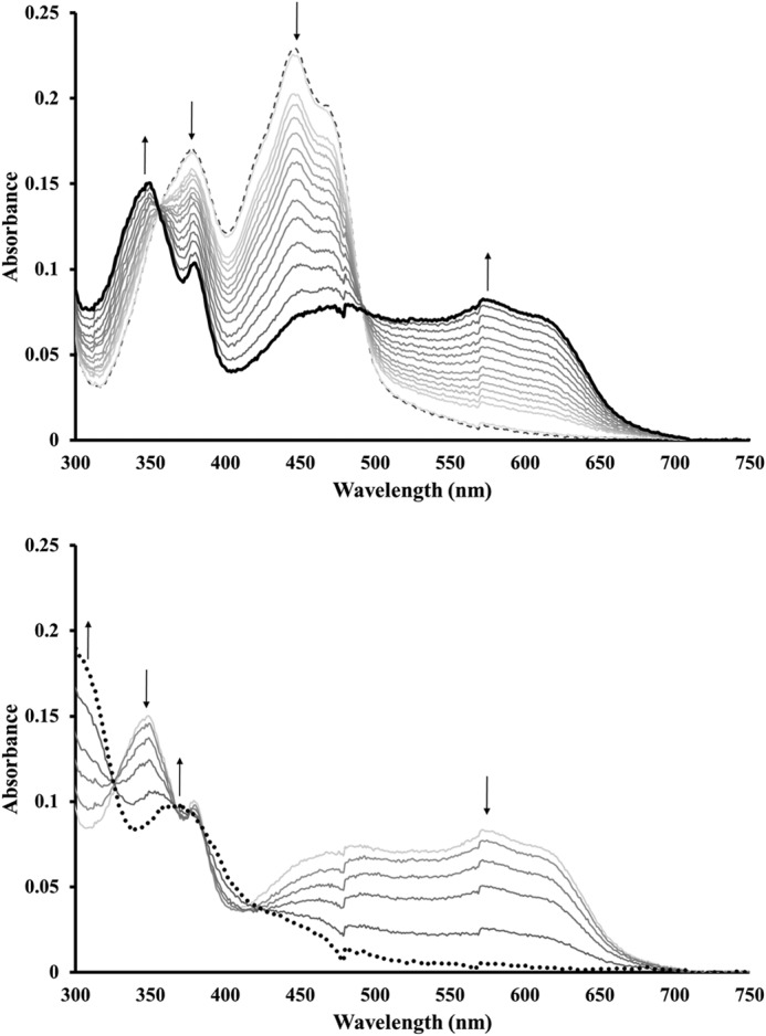
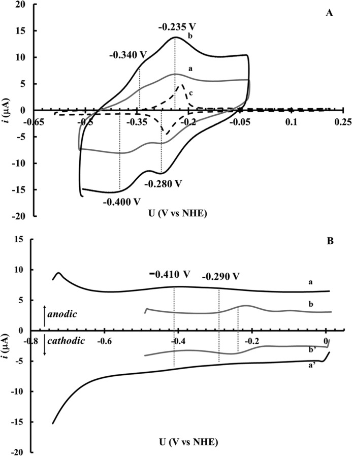
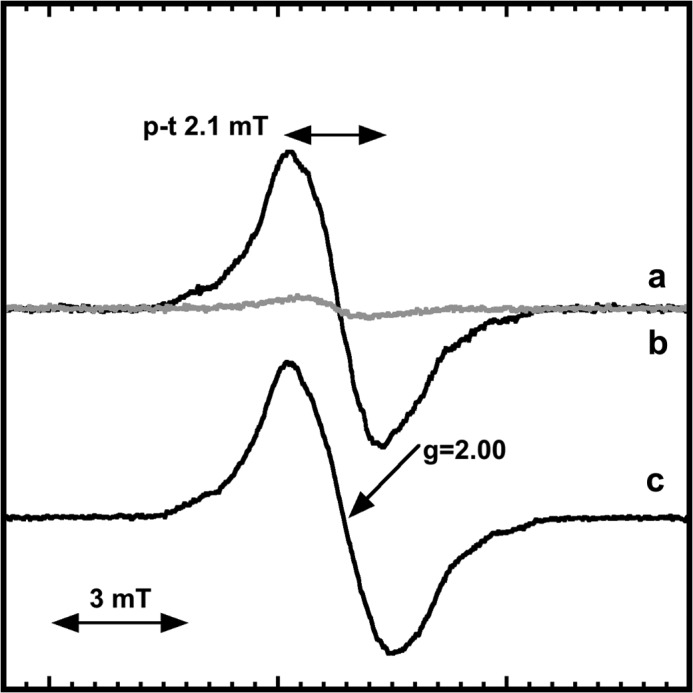
Ribonucleotide reductases (RNRs) catalyze the conversion of ribonucleotides to deoxyribonucleotides, and represent the only de novo pathway to provide DNA building blocks. Three different classes of RNR are known, denoted I-III. Class I RNRs are heteromeric proteins built up by α and β subunits and are further divided into different subclasses, partly based on the metal content of the β-subunit. In subclass Ib RNR the β-subunit is denoted NrdF, and harbors a manganese-tyrosyl radical cofactor. The generation of this cofactor is dependent on a flavodoxin-like maturase denoted NrdI, responsible for the formation of an active oxygen species suggested to be either a superoxide or a hydroperoxide. Herein we report on the magnetic properties of the manganese-tyrosyl radical cofactor of Bacillus anthracis NrdF and the redox properties of B. anthracis NrdI. The tyrosyl radical in NrdF is stabilized through its interaction with a ferromagnetically coupled manganese dimer. Moreover, we show through a combination of redox titration and protein electrochemistry that in contrast to hitherto characterized NrdIs, the B. anthracis NrdI is stable in its semiquinone form (NrdIsq) with a difference in electrochemical potential of ∼110 mV between the hydroquinone and semiquinone state. The under anaerobic conditions stable NrdIsq is fully capable of generating the oxidized, tyrosyl radical-containing form of Mn-NrdF when exposed to oxygen. This latter observation strongly supports that a superoxide radical is involved in the maturation mechanism, and contradicts the participation of a peroxide species. Additionally, EPR spectra on whole cells revealed that a significant fraction of NrdI resides in its semiquinone form in vivo, underscoring that NrdIsq is catalytically relevant.
Keywords: Bacillus; Cyclic Voltammetry; Free Radicals; Manganese; Maturase; NrdF; NrdI; Ribonucleotide Reductase; Semiquinone; Superoxide Ion.
© 2014 by The American Society for Biochemistry and Molecular Biology, Inc.
Figures
Similar articles
-
NrdH-redoxin protein mediates high enzyme activity in manganese-reconstituted ribonucleotide reductase from Bacillus anthracis.J Biol Chem. 2011 Sep 23;286(38):33053-60. doi: 10.1074/jbc.M111.278119. Epub 2011 Aug 6. J Biol Chem. 2011. PMID: 21832039 Free PMC article.
-
Mechanism of assembly of the dimanganese-tyrosyl radical cofactor of class Ib ribonucleotide reductase: enzymatic generation of superoxide is required for tyrosine oxidation via a Mn(III)Mn(IV) intermediate.J Am Chem Soc. 2013 Mar 13;135(10):4027-39. doi: 10.1021/ja312457t. Epub 2013 Feb 27. J Am Chem Soc. 2013. PMID: 23402532 Free PMC article.
-
Streptococcus sanguinis class Ib ribonucleotide reductase: high activity with both iron and manganese cofactors and structural insights.J Biol Chem. 2014 Feb 28;289(9):6259-72. doi: 10.1074/jbc.M113.533554. Epub 2013 Dec 31. J Biol Chem. 2014. PMID: 24381172 Free PMC article.
-
The manganese(IV)/iron(III) cofactor of Chlamydia trachomatis ribonucleotide reductase: structure, assembly, radical initiation, and evolution.Curr Opin Struct Biol. 2008 Dec;18(6):650-7. doi: 10.1016/j.sbi.2008.11.007. Epub 2008 Nov 27. Curr Opin Struct Biol. 2008. PMID: 19046875 Review.
-
The periodic table of ribonucleotide reductases.J Biol Chem. 2021 Oct;297(4):101137. doi: 10.1016/j.jbc.2021.101137. Epub 2021 Aug 27. J Biol Chem. 2021. PMID: 34461093 Free PMC article. Review.
Cited by
-
A Research-inspired biochemistry laboratory module-combining expression, purification, crystallization, structure-solving, and characterization of a flavodoxin-like protein.Biochem Mol Biol Educ. 2019 May;47(3):318-332. doi: 10.1002/bmb.21218. Epub 2019 Feb 11. Biochem Mol Biol Educ. 2019. PMID: 30742352 Free PMC article.
-
Chemical flexibility of heterobimetallic Mn/Fe cofactors: R2lox and R2c proteins.J Biol Chem. 2019 Nov 29;294(48):18372-18386. doi: 10.1074/jbc.RA119.010570. Epub 2019 Oct 7. J Biol Chem. 2019. PMID: 31591267 Free PMC article.
-
Metal-free ribonucleotide reduction powered by a DOPA radical in Mycoplasma pathogens.Nature. 2018 Nov;563(7731):416-420. doi: 10.1038/s41586-018-0653-6. Epub 2018 Oct 31. Nature. 2018. PMID: 30429545 Free PMC article.
-
Assembly of a heterodinuclear Mn/Fe cofactor is coupled to tyrosine-valine ether cross-link formation in the R2-like ligand-binding oxidase.J Biol Inorg Chem. 2019 Mar;24(2):211-221. doi: 10.1007/s00775-019-01639-4. Epub 2019 Jan 28. J Biol Inorg Chem. 2019. PMID: 30689052 Free PMC article.
-
Class Id ribonucleotide reductase utilizes a Mn2(IV,III) cofactor and undergoes large conformational changes on metal loading.J Biol Inorg Chem. 2019 Sep;24(6):863-877. doi: 10.1007/s00775-019-01697-8. Epub 2019 Aug 14. J Biol Inorg Chem. 2019. PMID: 31414238 Free PMC article.
References
-
- Nordlund P., Reichard P. (2006) Ribonucleotide Reductases. Annu. Rev. Biochem. 75, 681–706 - PubMed
-
- Schimpff-Weiland G., Follmann H. (1981) A new manganese-activated ribonucleotide reductase found in gram-positive bacteria. Biochem. Biophys. Res. Commun. 102, 1276–1282 - PubMed
-
- Willing A., Follmann H., Auling G. (1988) Nucleotide and thioredoxin specificity of the manganese ribonucleotide reductase from Brevibucterium ammoniagenes. Eur. J. Biochem. 175, 167–173 - PubMed
Publication types
MeSH terms
Substances
LinkOut - more resources
Full Text Sources
Other Literature Sources
