. 2014 Nov 1:123:225-35.
doi: 10.1016/j.colsurfb.2014.09.020. Epub 2014 Sep 19.

Bi-ligand surfaces with oriented and patterned protein for real-time tracking of cell migration

Affiliations

Bi-ligand surfaces with oriented and patterned protein for real-time tracking of cell migration

Varadraj N Vernekar et al. Colloids Surf B Biointerfaces. .

Abstract

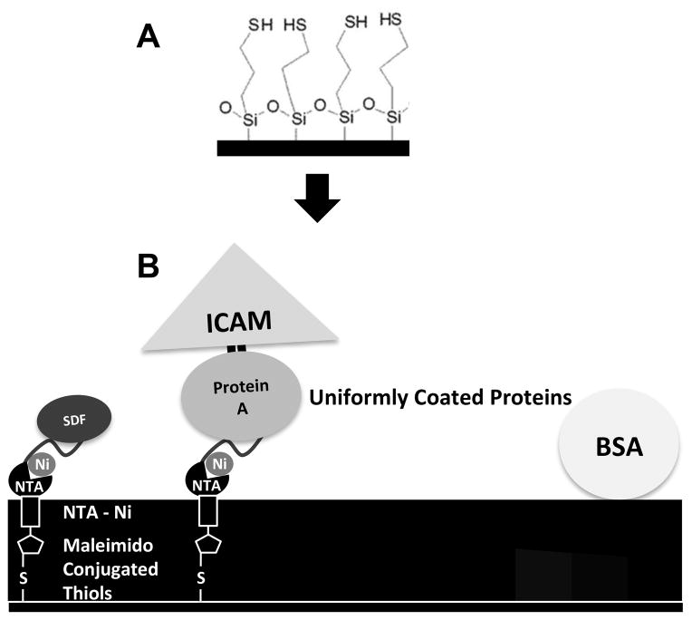
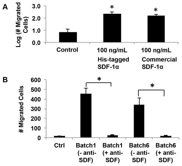
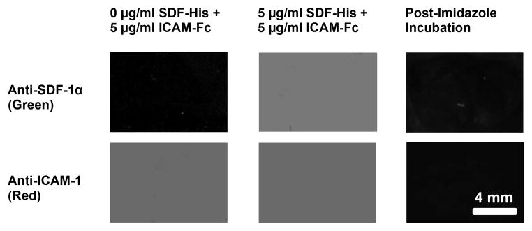
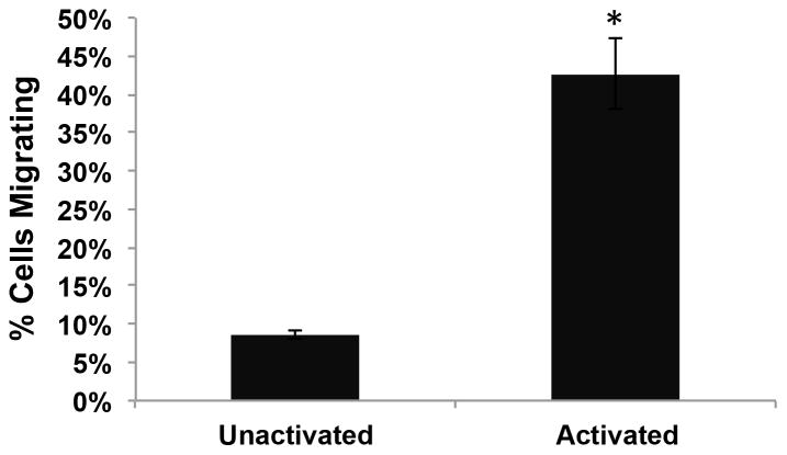
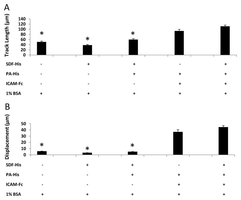
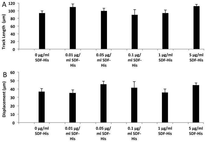
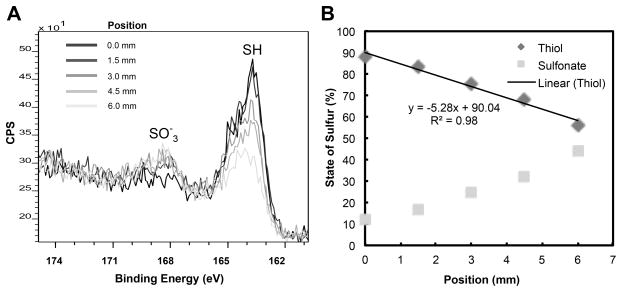
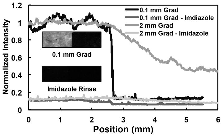
A bioactive platform for the quantitative observation of cell migration is presented by (1) presenting migration factors in a well-defined manner on 2-D substrates, and (2) enabling continuous cell tracking. Well-defined substrate presentation is achieved by correctly orienting immobilized proteins (chemokines and cell adhesion molecules), such that the active site is accessible to cell surface receptors. A thiol-terminated self-assembled monolayer on a silica slide was used as a base substrate for subsequent chemistry. The thiol-terminated surface was converted to an immobilized metal ion surface using a maleimido-nitrilotriacetic acid (NTA) cross-linker that bound Histidine-tagged recombinant proteins on the surface with uniform distribution and specific orientation. This platform was used to study the influence of surface-immobilized chemokine SDF-1α and cell adhesion molecule ICAM-1 on murine splenic B lymphocyte migration. While soluble SDF-1α induced trans-migration in a Boyden Chamber type chemotaxis assay, immobilized SDF-1α alone did not elicit significant surface-migration on our test-platform surface. Surface-immobilized cell adhesion protein, ICAM-1, in conjunction with activation enabled migration of this cell type on our surface. Controlled exposure to UV light was used to produce stable linear gradients of His-tagged recombinant SDF-1α co-immobilized with ICAM-1 following our surface chemistry approach. XPS and antibody staining showed defined gradients of outwardly oriented SDF-1α active sites. This test platform can be especially valuable for investigators interested in studying the influence of surface-immobilized factors on cell behavior and may also be used as a cell migration enabling platform for testing the effects of various diffusible agents.

Keywords: B lymphocyte; Cell adhesion molecule; Chemokine; Chemotaxis; Surface gradients; Surface immobilization.


Figures

Figure 1
Figure 1. Formation of uniformly coated protein surfaces
(A) Silica microscope slides were treated with MTS to form a uniform, thiol-terminated self-assembled monolayer on the surface. (B) A nitrilotriacetic acid (NTA)-containing cross-linker with a reactive maleimido group on one end (Maleimido-C3-NTA) enabled attachment of cross-linker to surface thiols. The NTA chelated Nickel. The Ni -- His-tag affinity enabled immobilization of both His-tagged SDF-1α (SDF-His) and His-tagged Protein A (PA-His), which were contacted on the surface at the same time for competitive binding. Following rinsing, cell adhesion molecule ICAM-1/Fc (ICAM-Fc) fusion chimera was introduced, which bound via affinity of the Fc region to Protein A. Once the proteins of interest were immobilized the surfaces were blocked with BSA. This process allowed the bioactive presentation of the active site (near N-terminus) of SDF-1α by attachment via C-terminus His-tag to Ni, and ICAM-1 by attachment via the Fc region to Protein A; and the formation of uniform surfaces of randomly distributed proteins.
Figure 2
Figure 2. In-house produced recombinant His-tagged SDF-1α is bioactive
(A) The bioactivity of His-tagged SDF-1α was verified via a trans-well migration assay, in which commercially available SDF-1α, His-tagged SDF-1α, or no chemokine control conditions were maintained in the lower chamber, whereas migrating cells were placed in the upper chamber separated by a thin porous membrane. The number of cells that crossed the membrane was quantified. A statistically significant increase in trans-migration of cells over the control case was observed for both in-house produced His-tagged SDF-1α and commercially purchased SDF-1α, and the latter two were not statistically distinguishable (One-way ANOVA, *P<0.05). (B) The specificity of His-tagged SDF-1α was confirmed, via an antibody neutralization assay, in which migration was tested with SDF-1α in the lower chamber that was equilibrated with or without anti-SDF antibody. Excess antibody neutralization (Ab:SDF-1α of 14:1) of two recombinant batches showed complete abolishment of chemotactic behavior to levels comparable to the control case (no chemokine).
Figure 3
Figure 3. Characterization of uniformly coated protein surfaces by fluorescent antibody binding
Fluorescent antibody binding to the surface was specific; insignificant monoclonal anti-SDF-1 antibody binding (green) was seen for a surface contacted with 0 μg/ml SDF-His (Column 1, Row 1), whereas significant antibody binding (green) was observed on surfaces contacted with 5 μg/ml SDF-His (Column 2, Row 1). Likewise, fluorescent antibody binding to the surface was reproducible; significant and comparable monoclonal anti-ICAM-1 antibody binding (red) was seen on different substrates contacted with the same 5 μg/ml ICAM-Fc concentration (Column 1, Row 2; and Column 2, Row 2). Finally, Significant abolishment of fluorescence (Column 3) upon Imidazole incubation of fluorescent antibody bound protein immobilized substrates (5 μg/ml ICAM-Fc + 5 μg/ml SDF-His contacted) demonstrated that the binding of His-tagged SDF-1α to the nickel coated surfaces was mostly specific, implying correct orientation of the protein with active site accessible.
Figure 4
Figure 4. Activation of cells is necessary for migration
The number of cells in 48–72 hour-old cultures that included 20μg/ml Anti-IgM and 20μg/ml Anti-CD40 in culture media exhibiting activated profiles and migratory behavior was significantly higher than in control cultures without these activating agents (Student’s t-test, *P = 0.002).
Figure 5
Figure 5. ICAM-1 is necessary for activated murine B lymphocyte surface migration
A one-way ANOVA on Ranks failed to show significant differences in cell migration on (10 μg/ml PA-His + 5 μg/ml ICAM-Fc + BSA) and (10 μg/ml PA-His + 5 μg/ml ICAM-Fc + 5 μg/ml SDF-His + BSA) contacted conditions for (A) track length and (B) displacement. On the other hand, displacement on BSA-contacted control surfaces and surfaces without ICAM-Fc was barely more than a cell body length and was significantly lower than the conditions containing ICAM-Fc (One way ANOVA of Ranks, * P ≤ 0.001). The same statistical differences were seen for track length, although the values for track length for these three non-ICAM-Fc conditions are greater than the characteristic cell body length; this is because the track length accrues even as a cell “dances” on the spot without much net displacement.
Figure 6
Figure 6. Immobilized SDF-1α does not significantly influence activated murine B lymphocyte surface migration beyond ICAM-1
Surfaces for each condition compared above were exposed to 10 μg/ml PA-His competitively with the specified concentrations of SDF-His followed by exposure to 5 μg/ml ICAM-Fc and blocking with 1% BSA. A one-way ANOVA of Ranks failed to show significant differences in (A) track length (* P = 0.098) and (B) displacement (* P = 0.296) between any of the concentrations of SDF-His for surfaces co-immobilized with ICAM-Fc via PA-His.
Figure 7
Figure 7. Characterization of the chemical surface gradient by XPS
(A) Graded exposure of the thiol surface to UV light by withdrawing a mask over 5 minutes across 6 mm resulted in opposing gradients of thiols and sulfonates. This is demonstrated by the progressively reducing SH peak and increasing SO3 peak (in the sulfur 2p region spectra, CPS = counts per second) as position on surface increases from 0 mm (end position of mask) to 6 mm (start position of mask). (B) The scaled area under the peaks was used to quantify the % of thiol and sulfonates with respect to the distance on surface that was exposed to UV.
Figure 8
Figure 8. Verification of specifically immobilized SDF-1α protein gradient
SDF-1α gradients (via binding of His-tagged SDF-1α to the Ni gradients) with varying slopes were formed by altering the removal speed of the UV mask and then stained with a fluorescent antibody against SDF-1α. The measured fluorescence intensities of (1) a steep gradient across 0.1 mm and (2) a shallow gradient across 2 mm were abolished with an imidazole rinse (which competes with the His-tag for affinity with Ni), demonstrating that SDF-1α binding to gradient is specific via the His-tag. This is visually demonstrated via the inset showing a scanned fluorescent image of the steep gradient before and after imidazole rinse.

References

    1. Singer S, Kupfer A. Annual Review of Cell Biology. 1986;2:337. - PubMed
    1. Thelen M. Nature Immunology. 2001;2:129. - PubMed
    1. Locascio A, Nieto MA. Current Opinion in Genetics & Development. 2001;11:464. - PubMed
    1. Condeelis J, Singer RH, Segall JE. Annu Rev Cell Dev Biol. 2005;21:695. - PubMed
    1. Chao DL, Ma L, Shen K. Nature Reviews Neuroscience. 2009;10:262. - PMC - PubMed

Publication types

Substances

LinkOut - more resources