Review
. 2015 Jan;53(1):143-59.
doi: 10.1002/dvg.22828. Epub 2014 Oct 6.

Quantitative and in toto imaging in ascidians: working toward an image-centric systems biology of chordate morphogenesis

Affiliations
Review

Quantitative and in toto imaging in ascidians: working toward an image-centric systems biology of chordate morphogenesis

Michael Veeman et al. Genesis. 2015 Jan.

Abstract

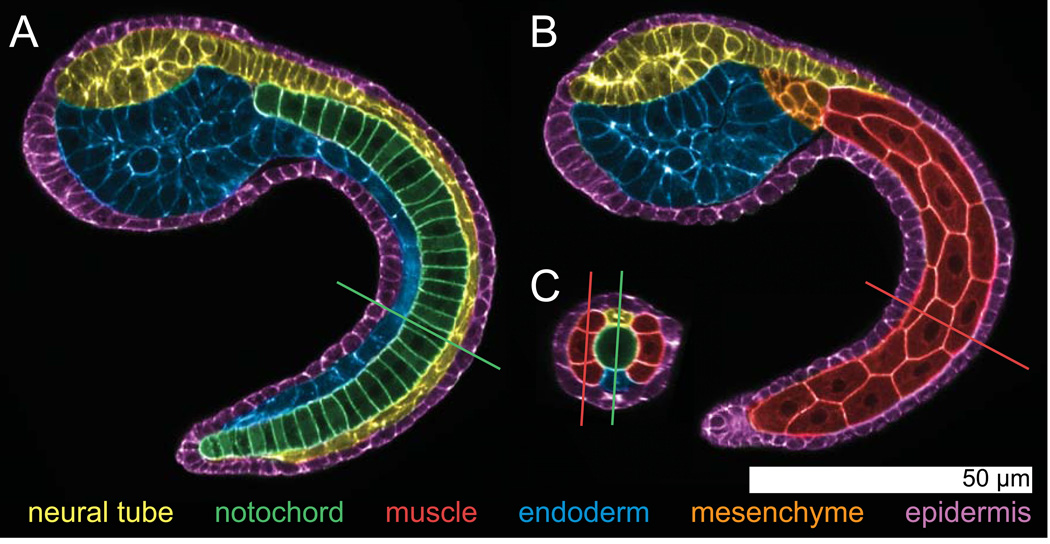
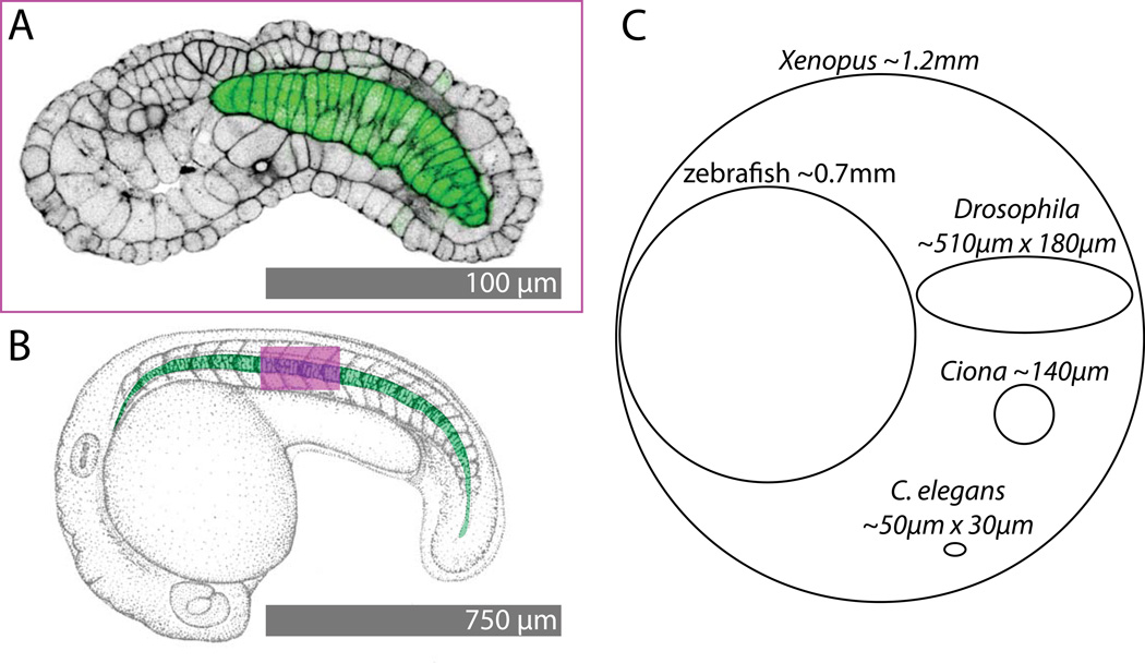
Developmental biology relies heavily on microscopy to image the finely controlled cell behaviors that drive embryonic development. Most embryos are large enough that a field of view with the resolution and magnification needed to resolve single cells will not span more than a small region of the embryo. Ascidian embryos, however, are sufficiently small that they can be imaged in toto with fine subcellular detail using conventional microscopes and objectives. Unlike other model organisms with particularly small embryos, ascidians have a chordate embryonic body plan that includes a notochord, hollow dorsal neural tube, heart primordium and numerous other anatomical details conserved with the vertebrates. Here we compare the size and anatomy of ascidian embryos with those of more traditional model organisms, and relate these features to the capabilities of both conventional and exotic imaging methods. We review the emergence of Ciona and related ascidian species as model organisms for a new era of image-based developmental systems biology. We conclude by discussing some important challenges in ascidian imaging and image analysis that remain to be solved.

Keywords: Ciona; ascidian; cell segmentation; in toto imaging; morphogenesis.


Figures

Figure 1
Figure 1. Anatomy of the Ciona intestinalis mid-tailbud embryo
Pseudocolored images from a confocal volume of a Hotta stage ~22 Ciona intestinalis embryo stained with Bodipy-FL-phallacidin to label the F-actin cytoskeleton, which is largely cortical during embryogenesis. The images were pseudocolored to highlight the 6 main tissues: epidermis, neural tube, notochord, tail muscle, mesenchyme and endoderm. The view in A has been resliced to follow the embryonic midline and shows the notochord in the center of the tail. The view in B is more superficial and shows the tail muscle cells and some mesenchymal cells. The hollow dorsal neural tube, endoderm and epidermis are evident in both A and B. C shows a cross section through the confocal volume at the position indicated by the lines in A and B. Conversely, the lines in C show the positions of the planes in A and B. The central notochord, dorsal neural tube and bilateral muscle cells are all evident in C. The confocal stack for these images was collected with a 40× 1.3NA objective in a single field of view on a standard confocal microscope (Zeiss LSM 700).
Figure 2
Figure 2. Size comparisons
A) Confocal image of phallacidin staining (black, contrast-reversed to match B) and a notochord-specific GFP transgene (green) in a stage ~19 Ciona intestinalis embryo. B) Pen and ink drawing of a zebrafish embryo at a comparable stage of early tail extension (modified from (Kimmel et al., 1995)). The notochord is pseudocolored in green. The magenta box shows the field of view of the Ciona image in A superimposed on the zebrafish embryo. C shows relative egg sizes for Xenopus (Brown, 2004), zebrafish (Kimmel et al., 1995), Drosophila (Markow et al., 2009), Ciona (Gregory and Veeman, 2013) and C. elegans (Begasse and Hyman, 2011).
Figure 3
Figure 3. Historical approaches
A) Hand-drawn image of a Ciona mid-tailbud embryo by Kowaleski (Kowalewski, 1866). The image shows 22 notochord cells instead of 40, and presents an overly regularized view of cell shape in the sensory vesicle and trunk endoderm, but the overall impression of the embryo’s chordate body plan is remarkably true to life. B) Hand-drawn image by Chabry of relatively normal gastrulation in an Ascidiella aspersa embryo where one of the two blastomeres had been killed with a fine needle at the two-cell stage (Chabry, 1887). C) Hand-drawn image by Conklin of the distribution of the yellow crescent material in the Styela egg that plays a key role in determining muscle cell fate (Conklin, 1905a).
Figure 4
Figure 4. Early in toto approaches to neural plate and notochord development
A,B) An early fate map of the Ciona neural plate through to early neural tube closure derived from histological sectioning and scanning EM of closely spaced timepoints of fixed embryos (Nicol and Meinertzhagen, 1988b). The blastomere numbering system used in these images is now obsolete, but the fundamental insight that the Ciona neural tube could be fate mapped with single cell resolution remains powerful. C,D,E) Pseudocolored confocal images of successive stages of notochord morphogenesis in Boltenia villosa (Munro and Odell, 2002b). These were among the first images to demonstrate how the entire ascidian embryo could be imaged by confocal microscopy to visualize fine details of cell and tissue morphology.
Figure 5
Figure 5. Quantitative approaches to cell shape and tissue architecture in toto
A–C are from (Tassy et al., 2006)). A) Computationally reconstructed cell shapes for an entire 32-cell stage Ciona embryo. One of the large B6.4 blastomeres and its much smaller B6.3 sibling are highlighted. The chart shows sibling cell volume asymmetries for several sib pairs. B) Rendered view of a 32-cell stage embryo highlighting the a6.5 blastomere in red and all of its immediate neighbors in yellow. C) a6.5 (red) shown next to its neighbor A6.2 (yellow) with touching surface areas highlighted in green. D) Epidermal cells segmented in toto in a mid-tailbud stage Ciona embryo (Nakamura et al., 2012).
Figure 6
Figure 6. Integrative uses of quantitative in toto imaging to study endoderm invagination and notochord tapering
A–C are from (Sherrard et al., 2011). A) Renderings of segmented embryos at successive time points during early gastrulation showing cell shape changes in the endoderm cells (yellow). B) Differential timing and extent of apical constriction and apical-basal shortening in endoderm cells (yellow) versus epidermal cells (red). C) Cartoon model of how early gastrulation is driven by a two step process involving first apical constriction and then subsequent apical-basal shortening. D-G are from (Veeman and Smith, 2012). D) Midvolume planes from confocal volumes of successive stages of notochord elongation. Segmented notochord cells are indicated with random pseudocolors. E) Normalized radius as a function of AP position for several stages of notochord elongation. Different timepoints (minutes after the end of notochord intercalation) are indicated with pseudocolors. F) Midvolume slice through a representative segmented Ciona notochord at the onset of mediolateral intercalation. Identified sibling pairs are connected with white lines. G) Notochord cell volumes as a function of AP position for three embryos segmented at the stage shown in F. Known sibling cells are connected by lines and colored according to the map in F.
Figure 7
Figure 7. Embryonic cell segmentation
A) Cartoon view of hypothetical cell membranes (green) and nuclei (magenta). B) Cell shapes can be represented in a vector format by lists of points connected by defined edges. C) Cell shapes can also be represented in a raster format by masks of pixels/voxels belonging to each cell. D) Raw confocal image of a section of Ciona tail. E) Preprocessing by membrane-enhancing CED filter. F) Hand-drawn seeds (green) for marker-assisted watershed segmentation. G) Watershed output with segmented cells labeled with random pseudocolors. H) Digitally ‘skinned’ embryo with the muscle/neural/endodermal strand cell layer flattened out to show the two blocks of muscle cells (large hexagonal/pentagonal cells) with neural cells between them (Abdollahian et al., 2011). One axis of this projection follows a curvilinear path along the AP axis. The other represents a radial axis around the embryonic mediolateral/dorsoventral midline.

References

    1. Abdollahian G, Veeman M, Smith W, Manjunath BS. A Curvicylindrical Coordinate System for the Visualization and Segmentation of the Ascidian Tail. Proc IEEE Int Symp Biomed Imaging. 2011:182–186. - PMC - PubMed
    1. Abitua PB, Wagner E, Navarrete IA, Levine M. Identification of a rudimentary neural crest in a non-vertebrate chordate. Nature. 2012;492:104–107. - PMC - PubMed
    1. Amos WB, White JG. How the confocal laser scanning microscope entered biological research. Biol Cell. 2003;95:335–342. - PubMed
    1. Begasse ML, Hyman AA. The first cell cycle of the Caenorhabditis elegans embryo: spatial and temporal control of an asymmetric cell division. Results Probl Cell Differ. 2011;53:109–133. - PubMed
    1. Brown DD. A tribute to the Xenopus laevis oocyte and egg. J Biol Chem. 2004;279:45291–45299. - PubMed

Publication types

LinkOut - more resources