. 2014 Sep 29:5:5072.
doi: 10.1038/ncomms6072.

XTACC3-XMAP215 association reveals an asymmetric interaction promoting microtubule elongation

Affiliations

XTACC3-XMAP215 association reveals an asymmetric interaction promoting microtubule elongation

Gulnahar B Mortuza et al. Nat Commun. .

Abstract

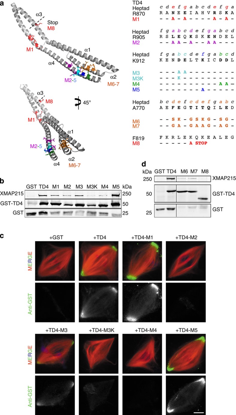
chTOG is a conserved microtubule polymerase that catalyses the addition of tubulin dimers to promote microtubule growth. chTOG interacts with TACC3, a member of the transforming acidic coiled-coil (TACC) family. Here we analyse their association using the Xenopus homologues, XTACC3 (TACC3) and XMAP215 (chTOG), dissecting the mechanism by which their interaction promotes microtubule elongation during spindle assembly. Using SAXS, we show that the TACC domain (TD) is an elongated structure that mediates the interaction with the C terminus of XMAP215. Our data suggest that one TD and two XMAP215 molecules associate to form a four-helix coiled-coil complex. A hybrid methods approach was used to define the precise regions of the TACC heptad repeat and the XMAP215 C terminus required for assembly and functioning of the complex. We show that XTACC3 can induce the recruitment of larger amounts of XMAP215 by increasing its local concentration, thereby promoting efficient microtubule elongation during mitosis.


Figures

Figure 1
Figure 1. Defining the minimal XMAP215 and XTACC3 binding domain and its effect on localization to the spindle poles and assembly.
(a) The chTOG/XMAP215 and XTACC3/Maskin domain architecture. Various C-terminal fragments of XMAP215 and XTACC3 were cloned and expressed for biophysical and functional analysis. (b) Representative images of spindles assembled in egg extracts containing GST, GST-TD4 and GST-TD5 at 150 nM. Samples were processed for immunofluorescence with an anti-GST antibody. GST-TD4 localized to the spindle poles, whereas GST-TD5 did not. (c) Representative images of spindles assembled in egg extracts containing GST or XMAP-Ct (15 μM). Samples were processed for immunofluorescence with anti-XTACC3 antibodies. Bipolar spindles also assemble in egg extracts containing XMAP-Ct, but their tubulin density and XTACC3 localization were reduced compared with controls. Images were taken with identical camera settings. In all cases, DNA is in blue, tubulin in red and GST in green. Scale bars, 10 μm (b,c). The experiment was repeated four times. (d) The interaction between TDs (TD4 and TD5) and XMAP-Ct was tested by SPR, and KD values were measured to show that XMAP-Ct has different binding affinities for TD4 and TD5. (e) SDS–PAGE showing pull-downs of TD4 using His-XMAP-Ct. Lane 1: reaction mixture; lanes 2–4: washes and lanes 5–6, elution. RU, response unit.
Figure 2
Figure 2. C-terminal binding region of XMAP-Ct is a bona fide coiled coil.
(a) Conformational chemical shift pattern by NMR of XMAP-Ct (pH 5.9 and 25 °C) as a function of the protein sequence suggests a helical propensity. Above: Cα (grey bars) and Cβ (white bars); below: comparison of the Cα conformational chemical shits of XMAP-Ct (white bars) and pCt (pH 6.1, 5 °C; grey bars). (b) A peptide derived from XMAP-Ct coiled-coil region was synthesized (pCt). Positions e and d of the heptad repeat were mutated to introduce either an opposite charge (pCt KD) or to break the hydrophobic interaction (pCt_LS). Scheme of a wheel diagram depicting the disposition of key residues in a simple two-helix coiled-coil interaction. A coiled-coil interaction of a more complex nature (trimer/tetramer) will also follow the same principle forming a hydrophobic core with the charged residues pointing outwards. (c) ITC and SPR binding curves showing TD4 interaction with pCt (red) and no interaction with pCt_KD (blue). The calculated KD values were 11 and 30 μM by ITC and SPR, respectively. (d) Western blot of pull-downs of CSF, XMAP-Ct and its mutants. Only XMAP-Ct and mutants of conserved charge (XMAP-Ct_KR and XMAP-Ct_LI) can pull down XTACC3, whereas by reversing the charge (XMAP-Ct_KD and XMAP-Ct_LS) mutants were unable to pull down XTACC3. The efficiency of the pull-down of the tagged proteins was verified by Coomassie blue staining. RU, response unit.
Figure 3
Figure 3. SAXS measurements and structure models of TD4.
(a) Pair distance distribution function p(r) for TD4 alone (black) or with pCt (red) are shown together in an ab initio GASBOR model. (b) A TD4 dimer model with and without P2 symmetry is composed of four helices (α1–α4). The models were manually constructed and were optimized using CORAL. The two models have also been turned 90° around two perpendicular axes to show different projections.
Figure 4
Figure 4. Mapping the XMAP-Ct binding site on TD4.
(a) SAXS structure of a TD4 monomer highlighting regions where mutations were performed in stick representation. Two regions on α4 were detected by NMR to change upon XMAP-Ct binding. (b) Western blot analysis of anti-GST pull-downs from egg extracts containing various GST fusion proteins corresponding to the different mutations on α4 of TD4. The blots were examined with anti-XMAP215 (upper lane) and anti-GST (middle and bottom lanes). GST-TD4, TD4-M1, TD4-M3 and TD4-M5 can pull down endogenous XMAP215. GST-TD4-M2 can also pull down XMAP215 but less efficiently. However, the mutants affecting the polarity of this heptad region (TD4-M3K and M4) strongly abrogated XMAP215–TD4 interaction. (c) Representative images of spindles assembled in egg extracts containing GST or the different GST-TD4 mutants as indicated. The samples were processed for immunofluorescence using the anti-GST antibody. Images were taken with identical camera settings. GST-TD4, TD4-M1, TD4-M3 and TD4-M5 localize at the spindle poles, whereas GST, -M2, -M3K and -M4 do not. DNA is in blue, tubulin in red and GST in green. Scale bar, 10 μm. (d) Western blot analysis of anti-GST pull-downs from egg extracts containing various GST fusion proteins corresponding to the different mutations on α2 and α3 of TD4, all of which affected the XMAP215–TD4 interaction. The blots were examined with anti-XMAP215 (upper lane) and anti-GST (middle and bottom lanes).
Figure 5
Figure 5. Asp922 and Asp923 are key TD4 residues on α4 for XTACC3–XMAP215 interaction and function.
(a) Representative images of spindles assembled in mock or XTACC3-depleted extract (Δ) containing GST, His-FL or His-FL-M4 as indicated. Samples were processed for immunofluorescence with anti-XTACC3 antibodies. His-FL-M4 did not localize to the spindle. (b) Quantification chart of spindle length and tubulin density of the different samples. Spindles assembled in XTACC3-depleted extracts (Δ) are 20% shorter, and their tubulin density is reduced by 30% compared with controls as previously described. Addition of His-FL to the depleted extract fully restored these parameters unlike that with His-FL-M4. Error bars represent the s.e.m. *P value <0.01, obtained applying a homoscedastic t-test. (c) Representative images of spindles assembled in mock or XTACC3-depleted extract (Δ) containing GST, His-FL or His-FL-M4 as indicated. Samples were processed for immunofluorescence with anti-XMAP215 antibodies. In XTACC3-depleted extracts supplemented with GST or His-FL-M4, XMAP215 localization to the spindle is strongly reduced. (d) Quantifications chart of XMAP215 localization normalized on tubulin density of the different samples. All values are the weighted mean of three independent experiments. Error bars represent the s.e.m. Spindles assembled in XTACC3-depleted extracts (Δ) supplemented with GST or His-FL-M4 display 40% less XMAP215 than controls. *P value <0.01, obtained applying a homoscedastic t-test. (e) Representative images of spindles assembled in extract containing GST, His-FL, His-FL-M4 as indicated. Samples were processed for immunofluorescence using the anti-XTACC3 antibody. (f) Chart showing the spindle length of the different assays. Spindles assembled in extracts supplemented with extra His-FL are longer than those assembled in extract containing GST or His-FL-M4. Error bars represent the s.e.m. P values ‘*’and ‘**’ are <0.05 and <0.01, respectively, obtained applying a homoscedastic t-test. In all cases, images were taken with identical camera settings. DNA is in blue, tubulin in red and XMAP215 in green. Scale bars, 10 μm.
Figure 6
Figure 6. A model depicting XTACC3–XMAP215 interaction and MT elongation.
(a) Model of the TD4–pCt interaction showing the SAXS structure of a TD monomer interaction with XMAP215 peptide (pCt) via the C-terminal coiled-coil association forming a four-helix bundle. The localization of the complex is steered by the TD. (b) A wheel diagram showing a possible association of the heptad repeats. The assembly would consist of a hetero-tetramer (X1 and X2- two molecules of XMAP215; T-TD, α2 and α4) forming an internal hydrophobic core, with the charged residues on the external surface. (c) A TD-directed recruitment of XMAP215 promotes MT growth whereby the TOG domains of XMAP215 add tubulin dimers processively.

References

    1. Holland A. J. & Cleveland D. W. Boveri revisited: chromosomal instability, aneuploidy and tumorigenesis. Nat. Rev. Mol. Cell. Biol. 10, 478–487 (2009). - PMC - PubMed
    1. Al-Bassam J., Larsen N. A., Hyman A. A. & Harrison S. C. Crystal structure of a TOG domain: conserved features of XMAP215/Dis1-family TOG domains and implications for tubulin binding. Structure 15, 355–362 (2007). - PubMed
    1. Brouhard G. J. et al.. XMAP215 is a processive microtubule polymerase. Cell 132, 79–88 (2008). - PMC - PubMed
    1. Reber S. B. et al.. XMAP215 activity sets spindle length by controlling the total mass of spindle microtubules. Nat. Cell. Biol. 15, 1116–1122 (2013). - PubMed
    1. Gergely F., Draviam V. M. & Raff J. W. The ch-TOG/XMAP215 protein is essential for spindle pole organization in human somatic cells. Genes Dev. 17, 336–341 (2003). - PMC - PubMed

Publication types

LinkOut - more resources