. 2015 Jan:11:449-58.
doi: 10.1016/j.actbio.2014.09.035. Epub 2014 Sep 28.

Engineering a vascularized collagen-β-tricalcium phosphate graft using an electrochemical approach

Affiliations

Engineering a vascularized collagen-β-tricalcium phosphate graft using an electrochemical approach

Yunqing Kang et al. Acta Biomater. 2015 Jan.

Abstract

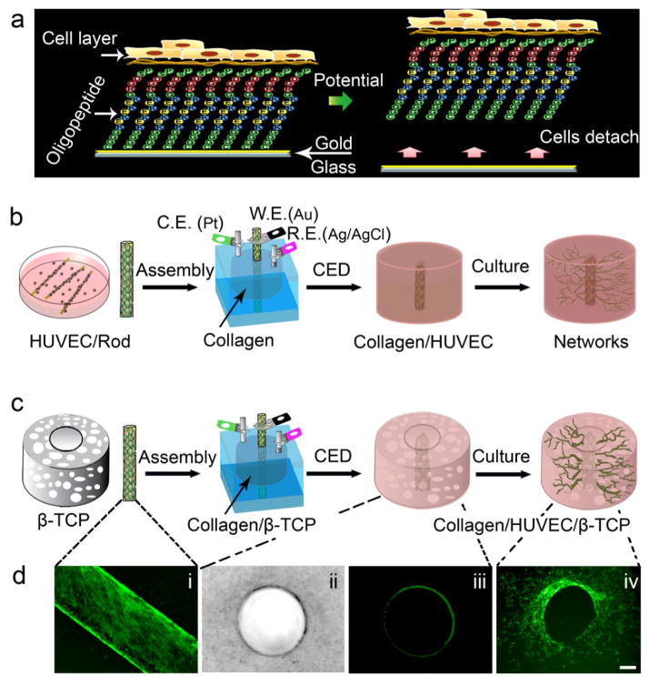
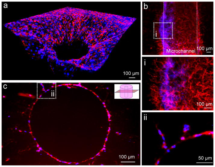
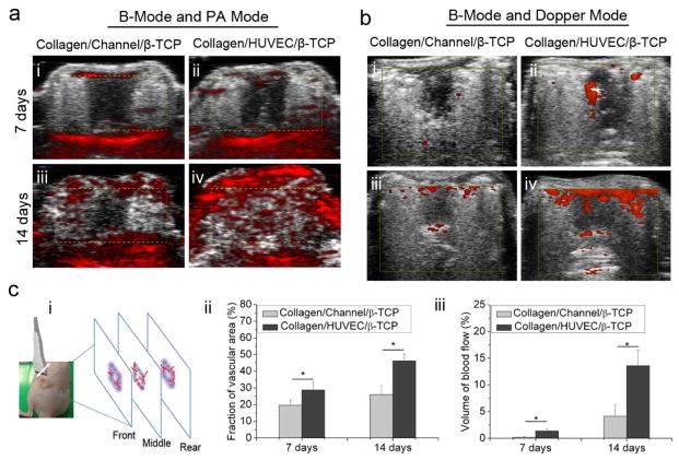
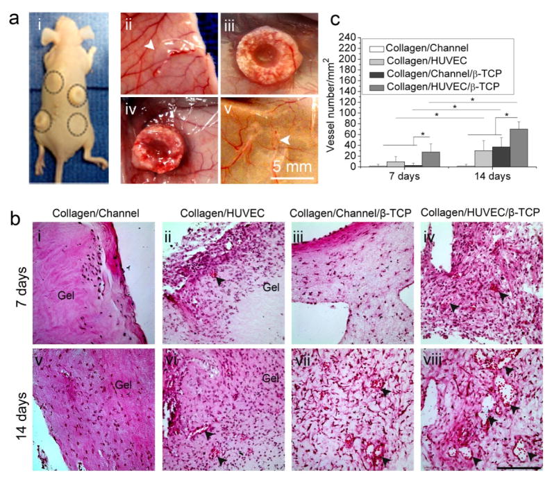
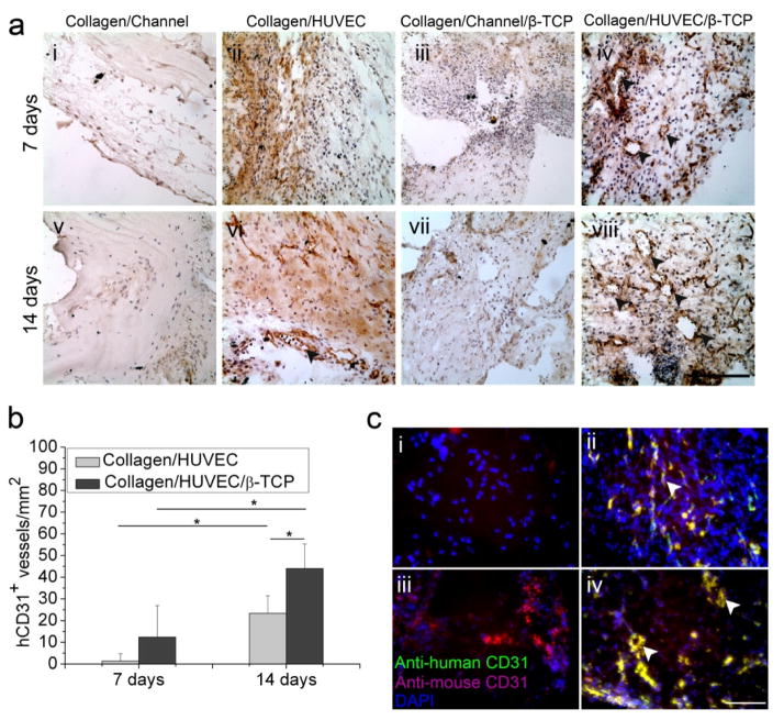
Vascularization of three-dimensional large synthetic grafts for tissue regeneration remains a significant challenge. Here we demonstrate an electrochemical approach, named the cell electrochemical detachment (CED) technique, to form an integral endothelium and use it to prevascularize a collagen-β-tricalcium phosphate (β-TCP) graft. The CED technique electrochemically detached an integral endothelium from a gold-coated glass rod to a collagen-infiltrated, channeled, macroporous β-TCP scaffold, forming an endothelium-lined microchannel containing graft upon removal of the rod. The in vitro results from static and perfusion culture showed that the endothelium robustly emanated microvascular sprouting and prevascularized the entire collagen/β-TCP integrated graft. The in vivo subcutaneous implantation studies showed that the prevascularized collagen/β-TCP grafts established blood flow originating from the endothelium-lined microchannel within a week, and the blood flow covered more areas in the graft over time. In addition, many blood vessels invaded the prevascularized collagen/β-TCP graft and the in vitro preformed microvascular networks anastomosed with the host vasculature, while collagen alone without the support of rigid ceramic scaffold showed less blood vessel invasion and anastomosis. These results suggest a promising strategy for effectively vascularizing large tissue-engineered grafts by integrating multiple hydrogel-based CED-engineered endothelium-lined microchannels into a rigid channeled macroporous scaffold.

Keywords: Collagen; Electrochemical; Microchannel; Vascularization; β-Tricalcium phosphate.


Figures

Fig. 1
Fig. 1
Fabrication of a microchannel in hydrogel-based or hydrogel-ceramic-based constructs. (a) A schematic shows the mechanism of the cell electrochemical detachment (CED) technique. (b) and (c) Schematic diagrams show the assembly procedures of a microchannel in a type I collagen hydrogel and in a collagen-β-TCP graft using the CED technique. (d) (i) a fluorescent image of HUVECs on an oligopeptide-coated rod, (ii) a bright-field image of a microchannel in collagen (top view), (iii) a fluorescent image of a microchannel in collagen (top view), and (iv) a fluorescent image of networks sprouting from the microchannel into surrounding area of the graft (top view)(Scale bar=100 μm).
Fig. 2
Fig. 2
Immunofluorescent images of human CD31 show network formation. (a) A reconstituted 3D confocal image shows the microcapillary networks extending to peripheral collagen matrix in a radial way. (b) A longitudinal view of a sprouting microchannel under a perfusion condition after 7 day incubation and these networks at a high magnification (i). (c) An immuofluorescent staining of CD31 on a paraffin section shows the capillary sprouting of HUVECs from the microchannel at a cross section view, and two sprouting branches at a higher magnification (ii). (Red: CD31; Blue: Nuclei)
Fig. 3
Fig. 3
Evaluations of the vascular volume and the blood flow in scaffolds from photoacoustic imaging and high-frequency Doppler ultrasound imaging systems. (a) B-mode and PA mode indicate the vascular volume in Collagen/Channel/β-TCP (i,iii) and Collagen/HUVEC/β-TCP (ii,iv) grafts at day 7 and 14 (yellow dotted box indicates the location of scaffold). (b) B-mode and Doppler mode indicate the blood flow density in Collagen/Channel/β-TCP (i,iii) and Collagen/HUVEC/β-TCP(ii,iv) at day 7 and 14 (yellow box indicates the location of scaffold). (c) Quantification of the vascular volume and blood flow density from PA and power Doppler images on day 7 and 14. (i) Transducer probe was moved above the sample and three slices images per sample were acquired for quantification. Color pixel density was used to express the vascular volume (ii) and perfused vascular density (blood flow) (iii) in the two groups. (*p<0.05, n=4).
Fig. 4
Fig. 4
Histological evaluation of angiogenesis of collagen-based and collagen-β-TCP-based grafts in vivo. (a) Macroscopic views of implants in the back of nude mice (i) and the four types of implants: Collagen/HUVEC (ii), Collagen/HUVEC/β-TCP (iii), Collagen/Channel/β-TCP (iv), and Collagen/Channel (v) on the skin at 14 days after implantation. White arrows show the collagen gels. (b) Representative images of H&E-stained sections from collagen-based and collagen-β-TCP-based grafts at day 7 and 14 (i-viii). Black arrowheads reveal the presence of blood vessels containing murine erythrocytes (Scale bar=100 μm). (c) Vessel density in the constructs over time. The number of vascular-like lumens was quantified for microvessel density. (*p<0.05, n=8).
Fig. 5
Fig. 5
Immunohistochemistry evaluation of angiogenesis and anastomosis in vivo. (a) Representative immunohistochemistry images of human-CD31 (hCD31) from Collagen/HUVEC and Collagen/HUVEC/β-TCP grafts at day 7 and 14 (i-viii) showed that the hCD31 positive microvessels contain murine erythrocytes (iv,viii) (black arrowheads) (Scale bar=100 μm). (b) The hCD31-positive expressing lumens containing murine erythrocytes were quantified by measuring their density (*p<0.05, n=8). (c) Immunofluorescent staining of human CD31 (green) and mouse CD31 (magenta) shows the anastomosed sites of preformed human capillaries with host vasculature (white arrows, yellow color). Four groups at day 14 are shown: (i) Collagen/Channel, (ii) Collagen/HUVEC, (iii) Collagen/Channel/β-TCP, and (iv) Collagen/HUVEC/β-TCP.

References

    1. Schek RM, Wilke EN, Hollister SJ, Krebsbach PH. Combined use of designed scaffolds and adenoviral gene therapy for skeletal tissue engineering. Biomaterials. 2006;27:1160–6. - PubMed
    1. Jarcho M. Calcium phosphate ceramics as hard tissue prosthetics. Clin Orthop Relat Res. 1981:259–78. - PubMed
    1. LeGeros RZ. Properties of osteoconductive biomaterials: calcium phosphates. Clin Orthop Relat Res. 2002:81–98. - PubMed
    1. Cuchiara MP, Gould DJ, McHale MK, Dickinson ME, West JL. Integration of Self-Assembled Microvascular Networks with Microfabricated PEG-Based Hydrogels. Adv Funct Mater. 2012;22:4511–8. - PMC - PubMed
    1. Chen YC, Lin RZ, Qi H, Yang Y, Bae H, Melero-Martin JM, et al. Functional Human Vascular Network Generated in Photocrosslinkable Gelatin Methacrylate Hydrogels. Adv Funct Mater. 2012;22:2027–39. - PMC - PubMed

Publication types

MeSH terms