. 2014 Oct 8;16(4):495-503.
doi: 10.1016/j.chom.2014.09.001. Epub 2014 Sep 25.

Discovery and characterization of gut microbiota decarboxylases that can produce the neurotransmitter tryptamine

Affiliations

Discovery and characterization of gut microbiota decarboxylases that can produce the neurotransmitter tryptamine

Brianna B Williams et al. Cell Host Microbe. .

Abstract

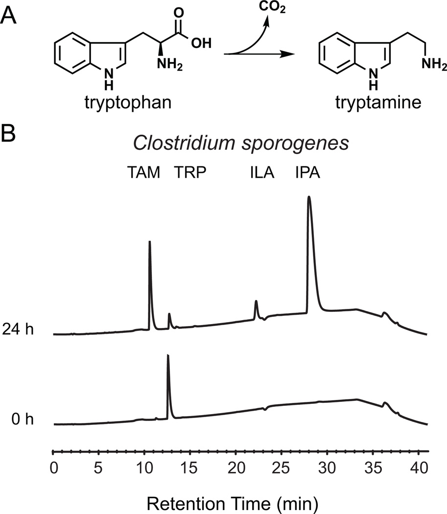
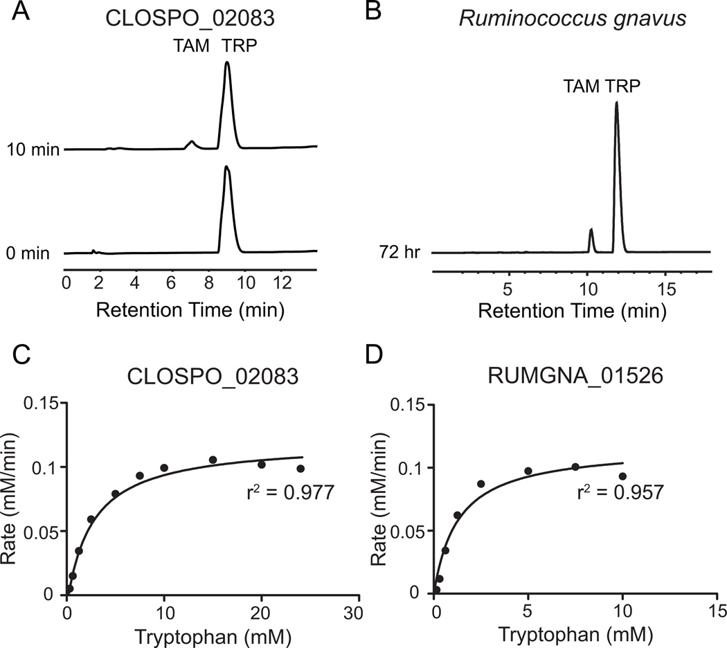
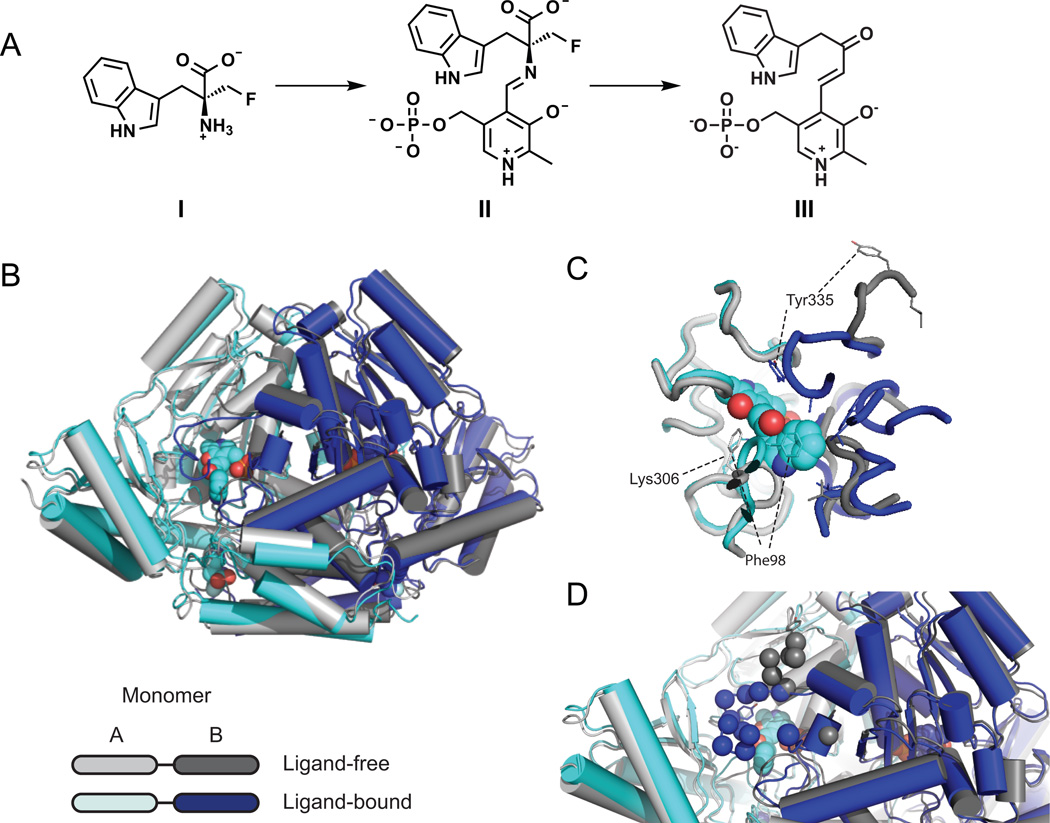
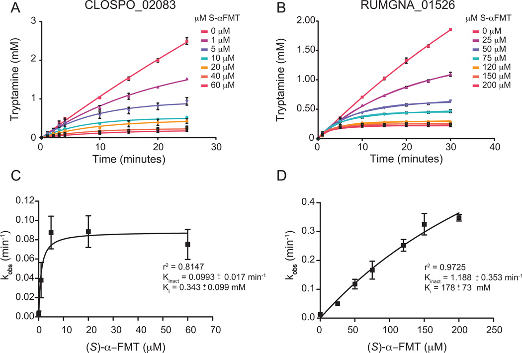
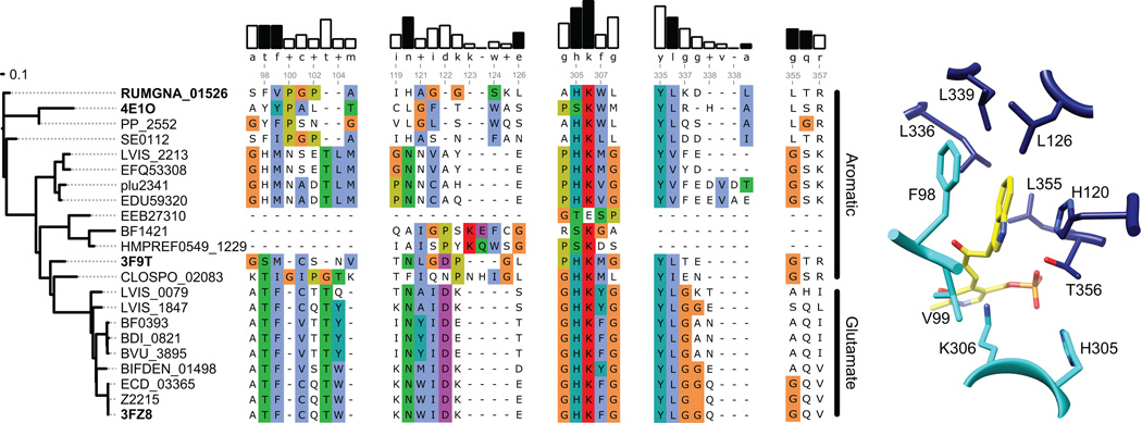
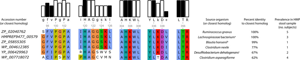
Several recent studies describe the influence of the gut microbiota on host brain and behavior. However, the mechanisms responsible for microbiota-nervous system interactions are largely unknown. Using a combination of genetics, biochemistry, and crystallography, we identify and characterize two phylogenetically distinct enzymes found in the human microbiome that decarboxylate tryptophan to form the β-arylamine neurotransmitter tryptamine. Although this enzymatic activity is exceedingly rare among bacteria more broadly, analysis of the Human Microbiome Project data demonstrate that at least 10% of the human population harbors at least one bacterium encoding a tryptophan decarboxylase in their gut community. Our results uncover a previously unrecognized enzymatic activity that can give rise to host-modulatory compounds and suggests a potential direct mechanism by which gut microbiota can influence host physiology, including behavior.


Figures

Figure 1
Figure 1. Tryptamine production by C. sporogenes
(A) The proteinogenic amino acid L-tryptophan is decarboxylated to tryptamine, a biogenic amine neurotransmitter, by the action of pyridoxal phosphate (PLP)-dependent decarboxylases. (B) Whole C. sporogenes were grown anaerobically in minimal media containing 5 g/L tryptophan, and clarified supernatant was analyzed by HPLC. C. sporogenes converts tryptophan (12.5 min) into tryptamine (TAM, 10.5 min), indole lactic acid (ILA, 22 min), and indole propionic acid (28 min). See also Figure S1 and S5.
Figure 2
Figure 2. CLOSPO_02083 and RUMGNA_01526 are Trp decarboxylases
(A) 100 nM purified CLOSPO_02083 was incubated with 2.5 mM tryptophan for 10 min and quenched with 1 volume MeOH; 100 µL of the reaction mixture was analyzed by HPLC. The HPLC trace shows the conversion of tryptophan (TRP, 9 min) to tryptamine (TAM, 7 min). (B) R. gnavus was grown anaerobically in minimal media containing 5 g/L tryptophan; 100 µL of the clarified supernatant was analyzed by HPLC. The HPLC trace shows the conversion of tryptophan (TRP, 12.5 min) to tryptamine (TAM, 10.9 min). Note: different HPLC methods were used for 2A and 2B. (C, D) Rate (mM tryptamine/min) vs. substrate concentration curves for tryptophan decarboxylation by (C) CLOSPO_02083 or (D) RUMGNA_01526. 100 nM enzyme was incubated with concentrations of tryptophan that varied from 0.15–24.5 mM. Error represents standard error of the mean. GraphPad was used to fit the Michaelis-Menten curve. See also Figure S2. Kinetic values are summarized in Figure S3.
Figure 3
Figure 3. Crystal structure of apo and ligand-bound RUMGNA_01526
(A) Schematic of proposed inhibitor mechanism: (S)-α-FMT (I) is converted to a PLP-(S)-α-FMT external aldimine intermediate (II), which is decarboxylated to a PLP-(S)-α-FMT Schiff base adduct (III). (B) Overlay of ligand-free (monomer A, light gray and monomer B, dark gray) and ligand-bound (monomer A, cyan and monomer B, blue) structures. In the active and allosteric sites, PLP-(S)-α-FMT and (S)-α-FMT (respectively) are shown in spheres. (C) Active site with PLP-(S)-α-FMT bound reveals a repositioning of Tyr335 and Phe98. In the ligand-bound structure, Lys306 is no longer covalently bound to PLP. (D) Upon engagement of (S)-α-FMT, residues 337–349 (dark blue spheres) fold over the active site, excluding solvent and forming critical interactions with the inhibitor. Dark gray spheres represent only ordered residues in apo structure. See also Figure S4 and S6.
Figure 4
Figure 4. Decarboxylase inhibition by (S)-α-FMT
Progress curve of tryptamine production by (A) CLOSPO_02083 and (B) RUMGNA_01526 in the presence of (A) 10 mM or (B) 2.5 mM tryptophan at various concentrations of inhibitor. Data were fit to the equation described in SI Materials and Methods to obtain kobs. (C, D) Plot of kobs vs [I]. CLOPSO_02083 is inhibited more potently by (S)-α-FMT than RUMGNA_01526 due to a higher binding affinity of the inhibitor. Error represents standard error of the mean. See also Figure S3.
Figure 5
Figure 5. Sequence and structural analysis of aromatic amino acid decarboxylases
(A) The dendrogram on the left shows the degree of sequence similarity between various decarboxylases. (B) Alignment of select amino acid decarboxylases are numbered according to the RUMGNA_01526 sequence. Four structural components of RUMGNA_01526 important for substrate binding are highlighted. The bars above consensus sequence show the degree of sequence conservation; residues from the RUMGNA_01526 structure that interact (black bars) or do not interact (white) with the tryptophan substrate are indicated. Residues in the sequence alignment are colored according to the Clustal color code (http://ekhidna.biocenter.helsinki.fi/pfam2/clustal_colours). (C) RUMGNA_10526 active site showing residues represented by black bars in (B).
Figure 6
Figure 6. Presence of tryptophan decarboxylase in the Human Microbiome Project Samples
Accession numbers of proteins of highest sequence identity to RUMGNA_01526 (ZP_02040762). BLAST percent identity was calculated for at least 100 amino acids. 15 subjects were found to contain homologs of the putative tryptophan decarboxylases. Of those, two contained 2 different homologs, and 13 contained one homolog. One subject harbored a gene with 93% identity to either ZP_0204072 from R. gnavus or HMPREF9477_00579 from Lachnospiraceae bacterium 2_1_58FAA. A sequence alignment is presented highlighting the residues identified by our structural analysis to be important for accommodating tryptophan (black bars). (a) Lachnospiraceae bacterium 2_1_58FAA (b) Blautia hansenii DSM 20583 (c) Desulfitobacterium dehalogenans ATCC 51507.

References

    1. Anderson GM. Quantitation of tryptophan metabolites in rat feces by thin-layer chromatography. Journal of chromatography. 1975;105:323–328. - PubMed
    1. Bell SM. Treatment with gentamicin monitored by serum antibiotic assay. The Medical journal of Australia. 1976;2:481–484. - PubMed
    1. Bhattacharjee MK, Snell EE. Pyridoxal 5'-phosphate-dependent histidine decarboxylase. Mechanism of inactivation by alpha-fluoromethylhistidine. The Journal of biological chemistry. 1990;265:6664–6668. - PubMed
    1. Bianchetti MG, Minder I, Beretta-Piccoli C, Meier A, Weidmann P. Effects of tyramine on blood pressure and plasma catecholamines in normal and hypertensive subjects. Klinische Wochenschrift. 1982;60:465–470. - PubMed
    1. Bischoff SC, Mailer R, Pabst O, Weier G, Sedlik W, Li Z, Chen JJ, Murphy DL, Gershon MD. Role of serotonin in intestinal inflammation: knockout of serotonin reuptake transporter exacerbates 2,4,6-trinitrobenzene sulfonic acid colitis in mice. American journal of physiology Gastrointestinal and liver physiology. 2009;296:G685–G695. - PubMed

Publication types

MeSH terms