. 2014 Oct 8;16(4):504-16.
doi: 10.1016/j.chom.2014.08.017. Epub 2014 Sep 25.

Epithelial IL-22RA1-mediated fucosylation promotes intestinal colonization resistance to an opportunistic pathogen

Affiliations

Epithelial IL-22RA1-mediated fucosylation promotes intestinal colonization resistance to an opportunistic pathogen

Tu Anh N Pham et al. Cell Host Microbe. .

Abstract

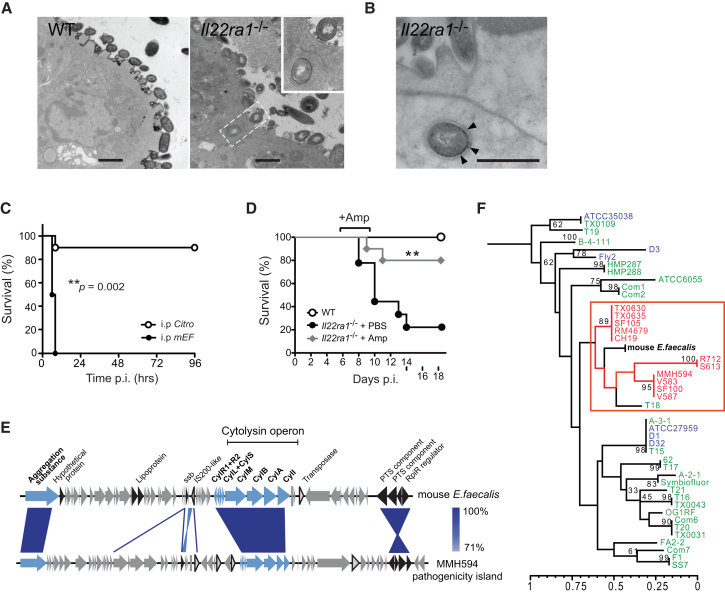
Our intestinal microbiota harbors a diverse microbial community, often containing opportunistic bacteria with virulence potential. However, mutualistic host-microbial interactions prevent disease by opportunistic pathogens through poorly understood mechanisms. We show that the epithelial interleukin-22 receptor IL-22RA1 protects against lethal Citrobacter rodentium infection and chemical-induced colitis by promoting colonization resistance against an intestinal opportunistic bacterium, Enterococcus faecalis. Susceptibility of Il22ra1(-/-) mice to C. rodentium was associated with preferential expansion and epithelial translocation of pathogenic E. faecalis during severe microbial dysbiosis and was ameloriated with antibiotics active against E. faecalis. RNA sequencing analyses of primary colonic organoids showed that IL-22RA1 signaling promotes intestinal fucosylation via induction of the fucosyltransferase Fut2. Additionally, administration of fucosylated oligosaccharides to C. rodentium-challenged Il22ra1(-/-) mice attenuated infection and promoted E. faecalis colonization resistance by restoring the diversity of anaerobic commensal symbionts. These results support a model whereby IL-22RA1 enhances host-microbiota mutualism to limit detrimental overcolonization by opportunistic pathogens.


Figures

None
Graphical abstract
Figure 1
Figure 1
IL-22RA1 Limits Systemic Bacterial Dissemination during Intestinal Disease (A) C. rodentium cfus in systemic organs. (B) Serum anti-C. rodentium EspA IgG titer. (C and D) TNF-α and IL-6 cytokines (C) and E. faecalis cfus (D) in systemic organs of infected WT and Il22ra1−/− mice. Shown are mean ± SEM in five independent infections (n = 4–6 each); ∗∗∗p < 0.0001. (E and F) Profile of systemic bacterial isolates from (E) WT (n = 10) and Il22ra1−/− mice infected with C. rodentium (n = 28) and (F) DSS-treated WT and Il22ra1−/− mice (n = 8–10). Numbers indicate total number of isolates assigned to a particular bacterial taxon in 3–8 independent experiments. See also Figure S2 and Tables S1 and S2.
Figure 2
Figure 2
A Pathogenic Enterococcus faecalis Isolate Harbors Virulence Factors, Translocates Intracellularly in Il22ra1-Deficient Mice, and Causes Lethal Septicemia (A) Transmission electron micrographs of C. rodentium-infected WT and Il22ra1−/− mice cecal tissues, showing translocation of a coccal bacterium. (B) Enterococcus-specific immunogold labeling (arrows) of an intracellular bacterium. Scale bar, 500 nm. (C) Survival of WT mice infected i.p. with either C. rodentium or E. faecalis (n = 10–12). (D) Survival C. rodentium-infected WT mice and Il22ra1−/− mice given i.p. ampicillin or PBS. Log-rank p value of two independent experiments (n = 5 each). (E) Schematic alignment of the cytolysin operon and flanking genes of mEF and the E. faecalis pathogenicity island. Blue, cytolysin and aggregation substance; black, other pathogenicity island-associated genes; triangles, transposase and IS elements. rpiR, family transcriptional regulator; PTS, phosphotransferase system; CylI, cytolysin immunity protein; CylA, cytolysin activator; CylB, cytolysin B ABC-type transporter; CylM, cytolysin subunit modifier; CylL+S, cytolysin subunits L–S; CylR1+R2, cytolysin regulators R1 and R2. (F) MLST-based phylogenetic relationship between mEF and nosocomial (red), community/probiotic (green), and animal (blue) E. faecalis strains. See also Figure S4 and Table S3.
Figure 3
Figure 3
IL-22RA1 Signaling Restricts Intestinal Dysbiosis and Enterococcus faecalis Overcolonization during C. rodentium Infection (A) Shannon diversity index of the fecal microbiota of WT and Il22ra1−/− mice (mean ± SEM) before C. rodentium infection (day 0) and at day 9 p.i. p < 0.05, ∗∗p < 0.001, ∗∗∗p < 0.0001. (B) Cluster dendogram of the microbial community structures of WT and Il22ra1−/− (KO) fecal and cecal microbiota (n = 3) before infection (day 0) and at day 9 p.i. at the operational taxonomic unit (OTU) level. Bar graphs represent proportional abundance. (C) Immunofluorescence of C. rodentium and Enterococcus spp. in the cecum of infected WT and Il22ra1−/− mice. DAPI, cell nuclei. Scale bar, 50 μm. (D) Fecal Enterococcus shedding of WT and Il22ra1−/− mice (n = 6–15) by selective plating, showing significant overgrowth during C. rodentium infection. (E) Selective plating and 16S rRNA sequencing profile of enterococci in the feces and cecal mucosa of infected WT (n = 15) and Il22ra1−/− mice (n = 40) in five independent experiments, showing preferential expansion of E. faecalis (red) relative to other intestinal enterococci (E. gallinarum, gray). Shown are numbers of Enterococcus sequences matched to a species ID. See also Figure S5.
Figure 4
Figure 4
RegIIIγ−/− Mice Did Not Show Enhanced Susceptibility to C. rodentium (A–E) WT and RegIIIγ−/− mice were infected orally with 109C. rodentium. (A) Shedding of C. rodentium and (B) E. faecalis over the course of infection. Bacterial load of (C) C. rodentium and (D) E. faecalis in the cecal lumen and mucosal tissues, determined by selective plating. (E) Cecal histopathology of C. rodentium-infected WT and RegIIIγ−/− mice at day 9 p.i., showing similar submucosal edema (asterisks) and inflammatory infiltrates (arrows). Scale bar, 100 μm. Results are from three independent experiments (n = 6–8 each).
Figure 5
Figure 5
RNA-Seq of IL-22RA1 Signaling in Colonic Epithelial Organoids Identifies Factors Associated with Host-Microbe Interactions (A) LacZ staining of WT and Il22ra1−/− organoids (scale bar, 100 μm) (left) and transmission electron microscopy of an Il22ra1−/− organoid with microvilli (Mv), tight junctions (arrowheads), and goblet cell (G) (right; scale bar, 1 μm). (B) RNA-seq transcriptomes of colonic organoids from wild-type (WT) and Il22ra1−/− (KO) mice (n = 4) treated with IL-22 or untreated (Ctrl) in technical replicates, showing hierarchical clustering. Colors indicate levels of correlation (low, light green-yellow; high, dark green-blue). (C) Enriched biological processes among upregulated (orange) and downregulated (blue) genes downstream of IL-22RA1 signaling, with selected genes associated with glycosylation. (D) Stat3-focused interaction network of genes induced by IL-22RA1 signaling (p < 10−12), 18 of which are associated with human susceptibility to chronic inflammatory diseases (gold). Lines indicate protein-protein interactions (dark/light blue), coexpression (black), colocalization (gray), and shared protein domains (cyan). See also Figure S6.
Figure 6
Figure 6
IL-22RA1-Mediated Fucosylated Glycan Expression Contributes to Host Defense (A) Cecal Fut2 transcripts of WT and Il22ra1−/− mice (n = 9–12) at day 9 p.i. with C. rodentium. (B) Immunofluorescence staining of WT and Il22ra1−/− cecal tissues with Lotus tetragonolobus lectin (green). (C–E) WT mice and groups of Il22ra1−/− littermates were treated orally with 2′-fucosyllactose (2′FL), blood group H disaccharide (or H antigen; H-Ag), lactose, or PBS between days 5 and 8 p.i. during C. rodentium infection. Shown are (C) survival (left) and weight loss (right), (D) histopathology (scale bar, 200 μm), and (E) total number of cLP leukocytes at day 9 p.i. (F and G) qPCR of (F) Th1 cytokines (IFNγ, TNF-α, IL-1β, IL-6) and (G) Th17-associated cytokines (IL-17a, IL-17f, IL-21, IL-22) in the cecal tissues at day 9 p.i.
Figure 7
Figure 7
Fucosylated Glycan Expression Enhances Colonization Resistance to E. faecalis (A–D) C. rodentium-infected WT mice and groups of Il22ra1−/− littermates were treated orally with 2′-fucosyllactose (2′FL) or PBS. Shown are (A) C. rodentium shedding, (B) E. faecalis cfus in the colonic lumen and mucosa (gray area, detection limit), (C) E. faecalis cfus in the liver and spleen, and (D) serum IL-6 and TNF-α levels at day 9 p.i. Mean ± SEM of data from three independent experiments (n = 3–5 each); p < 0.05, ∗∗p < 0.01, ∗∗∗p < 0.0001. (E) Shannon diversity index of the fecal microbiota before infection (day 0) and at day 9 p.i. (F) Cluster dendogram of the fecal microbiota community structures of 2′FL-treated Il22ra1−/− mice and PBS-treated controls (Ctrl). Bar graphs represent proportional abundance.

Comment in

References

    1. Arias C.A., Murray B.E. The rise of the Enterococcus: beyond vancomycin resistance. Nat. Rev. Microbiol. 2012;10:266–278. - PMC - PubMed
    1. Aujla S.J., Chan Y.R., Zheng M., Fei M., Askew D.J., Pociask D.A., Reinhart T.A., McAllister F., Edeal J., Gaus K. IL-22 mediates mucosal host defense against Gram-negative bacterial pneumonia. Nat. Med. 2008;14:275–281. - PMC - PubMed
    1. Becker D.J., Lowe J.B. Fucose: biosynthesis and biological function in mammals. Glycobiology. 2003;13:41R–53R. - PubMed
    1. Behnsen J., Jellbauer S., Wong C.P., Edwards R.A., George M.D., Ouyang W., Raffatellu M. The cytokine IL-22 promotes pathogen colonization by suppressing related commensal bacteria. Immunity. 2014;40:262–273. - PMC - PubMed
    1. Benjamin J.L., Sumpter R., Jr., Levine B., Hooper L.V. Intestinal epithelial autophagy is essential for host defense against invasive bacteria. Cell Host Microbe. 2013;13:723–734. - PMC - PubMed

Publication types

MeSH terms

LinkOut - more resources