Review
. 2014 Nov;12(11):739-49.
doi: 10.1038/nrmicro3346. Epub 2014 Sep 29.

The sweet spot: defining virus-sialic acid interactions

Affiliations
Review

The sweet spot: defining virus-sialic acid interactions

Jennifer E Stencel-Baerenwald et al. Nat Rev Microbiol. 2014 Nov.

Abstract

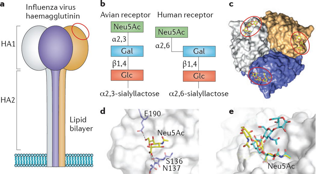
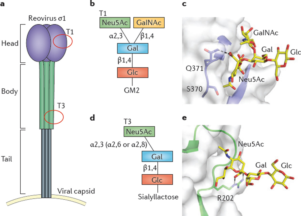
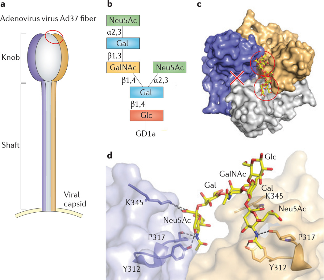
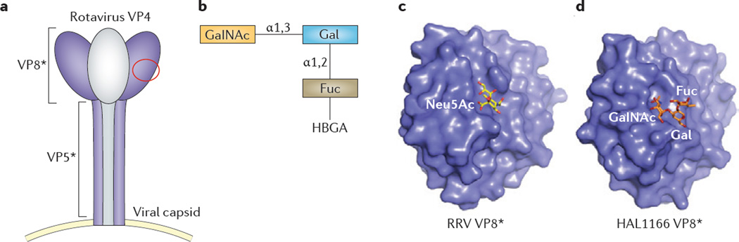
Viral infections are initiated by attachment of the virus to host cell surface receptors, including sialic acid-containing glycans. It is now possible to rapidly identify specific glycan receptors using glycan array screening, to define atomic-level structures of virus-glycan complexes and to alter the glycan-binding site to determine the function of glycan engagement in viral disease. This Review highlights general principles of virus-glycan interactions and provides specific examples of sialic acid binding by viruses with stalk-like attachment proteins, including influenza virus, reovirus, adenovirus and rotavirus. Understanding virus-glycan interactions is essential to combating viral infections and designing improved viral vectors for therapeutic applications.


Figures

Figure 1
Figure 1. Sialic acid types and glycosidic linkage
a | Sialic acids are none-carbon monosaccharide derivatives of neuraminic acid. The two most common sialic acids are N-acetyl neuraminic acid (Neu5Ac) and N-glycolylneuraminic acid (Neu5Gc). The C5 carbon in Neu5Ac is modified with an N-acetyl group, which can be further hydroxylated to form Neu5Gc. The hydroxyl groups at C4, C7, C8 and C9 are subject to various modifications (not shown). Common constituents include O-acetyl, O-sulphate, O-lactyl, O-methyl and O-phosphate groups. b | Sialic acids are attached to carbohydrate chains on glycoproteins and glycolipids via different glycosidic linkages. The most common linkage types are α2,3-linkage to a galactose residue, α2,6-linkage to a galactose moiety or to an N-acetylgalactosamine moiety, and α2,8-linkage to another sialic acid moiety on a glycan.
Figure 2
Figure 2. Influenza virus binding to differentially linked sialic acids
a | Schematic of the trimeric influenza virus haemagglutinin, with the monomers depicted in purple, orange and grey. Haemagglutinin is a transmembrane protein that is composed of the globular HA1 domain and the stalk-like HA2 domain. Each HA1 domain in the trimer binds to sialic acid (commonly N-acetyl neuraminic acid (Neu5Ac)), and the binding site is indicated in one monomer with a red circle. b | Avian influenza viruses preferentially bind to host cell receptors that contain α2,3-linked sialic acid, and human-adapted viruses bind to receptors that contain α2,6-linked sialic acid moieties. Schematics of an example of an avian influenza virus receptor (α2,3-sialyllactose) and a human influenza virus receptor (α2,6-siallylactose) are shown. Glucose (Glc), galactose (Gal) and Neu5Ac are depicted as blocks. c | Surface representation of trimeric haemagglutinin (monomers are shown in purple, orange and grey) in complex with Neu5Ac (in yellow as a stick representation) (Protein Data Bank (PDB) accession 1HGG). Red circles indicate the glycan-binding site. d | Close-up view of the glycan-binding site of haemagglutinin. Selected crucial contacts between the haemagglutinin residues Ser136, Asn137 and Glu190 (purple) and Neu5Ac (yellow) are highlighted (grey dashes). The glycan receptors are shown in stick representation, with oxygen atoms in red and nitrogen atoms in blue. e | Superposition of an avian influenza virus haemagglutinin in complex with α2,3-sialyllactosamine (yellow) (PDB accession 2WR2) and the human receptor α2,6-sialyllactosamine (cyan) (PDB accession 2WR7). The avian receptor generally has a linear conformation, whereas the human receptor is more flexible and has an umbrella-like topology.
Figure 3
Figure 3. T1 and T3 reovirus σr1 proteins differentially bind to sialylated glycans
a | Schematic showing the reovirus attachment protein σ 1, which is atrime ric fibre that is composed of three structurally distinct domains: the head (purple), body (green) and tail (grey). The glycan-binding sites of serotypes 1 (T1) and 3 (T3) are located in different domains of σ 1 (indicated with red circles). b | T1 reovirus binds to the GM2 glycan, which is composed of glucose (Glc) and galactose (Gal), with an α2,3-linked N-acetyl neuraminic acid (Neu5Ac), and β1,4-linked N-acetylgalactosamine (GalNAc). c | Close-up view of the T1 reovirus-GM2 interaction (Protein Data Bank (PDB) accession 4GU3). The protein surface is depicted in white, and the glycan-binding site is shown as a ribbon tracing in blue. Ser370 and Gln371 are crucial residues that are involved in GM2 binding and are shown in stick representation. The glycan receptor is shown in stick representation (yellow), with oxygen (red) and nitrogen atoms (blue). d | T3 reovirus binds to α2,3-, α2,6-, and α2,8-linked sialylated glycans. e | Close-up view of T3 reovirus σ 1 in complex with α2,3-linked sialyllactose (PDB accession 3S6X). The protein surface is shown with the glycan-binding site depicted as a ribbon tracing in green. Arg202, which is required for the virus–sialic acid interaction, is shown in stick representation. Contacts between viral residues and sialic acid are depicted as grey dashes.
Figure 4
Figure 4. Interaction between adenovirus 37 and glycan
a | Schematic representation of the trimeric adenovirus 37 (Ad37)fibre, which is the viral attachment, protein that binds to the GD1a glycan. The monomers are depicted in purple, orange and grey. The glycan-binding site is located in the knob domain of two monomers (indicated by a red circle). b | Schematic showing the GD1a glycan, which is the Ad37 glycan receptor on host cells. GD1a is composed of glucose (Glc), galactose (Gal), a terminal α2,3-linked sialic acid and N-acetylgalactosamine (GalNAc), depicted as blocks. c | Surface representation of a top view of the Ad37 fibre knob in complex with the GD1a glycan (Protein Data Bank (PDB) accession 3N0I). The monomers are coloured as in part a, and the GD1a glycan is shown in stick representation (in yellow, with oxygen atoms in red and nitrogen atoms in blue). The two N-acetyl neuraminic acid (Neu5Ac)-binding sites that are occupied by the GD1a glycan are marked with red circles, and the third potential binding site (X) remains unoccupied. d | Surface representation of two knob monomers bound to the GD1a glycan, which is shown in stick representation (oxygen and nitrogen atoms as in part c). The interaction between the Ad37 fibre knob and sialic acid is mediated by several interactions (depicted as grey dashes), including a salt bridge between Lys345 and the Neu5Ac carboxylate, and hydrogen bonds between residues Tyr312 and Pro317 of the Ad37 knob and the N-acetyl chain of Neu5Ac of GD1a.
Figure 5
Figure 5. The VP8* domain of rotavirus VP4 differentially engages glycans
a | The schematic depicts the rotavirus outer-capsid protein VP4, which is composed of VP5* and VP8*. The protein is a trimer, but only two of the three monomers are visible in some structures, and hence the third monomer is depicted in grey. The VP8* subunit binds to glycans (the binding site is indicated by the red circle), whereas the VP5* subunit facilitates membrane penetration. b | The glycan ligand for the human HAL1166 rotavirus, human blood group antigen (HBGA), comprises N-acetylgalactosa-mine (GalNAc), galactose (Gal) and fucose (Fuc). c | The crystal structure of rhesus rotavirus (RRV) VP8* in complex with N-acetyl neuraminic acid (Neu5Ac) (Protein Data Bank (PDB) accession 1KQR). The protein surface is shown in purple, and Neu5Ac is depicted in stick representation (yellow carbon with red oxygen atoms and blue nitrogen atoms). d | The crystal structure of human rotavirus strain HAL1166 VP8* (purple) in complex with HBGA (with orange carbons) (PDB accession 4DRV). The glycan receptors are shown as orange sticks with red oxygen atoms and blue nitrogen atoms. The HAL1166 VP8* binds to a completely different glycan at the same position at which RRV VP8* engages Neu5Ac.

Similar articles

Cited by

References

    1. Barton ES, Connolly JL, Forrest JC, Chappell JD, Dermody TS. Utilization of sialic acid as a coreceptor enhances reovirus attachment by multistep adhesion strengthening. J. Biol. Chem. 2001;276:2200–2211. - PubMed
    1. Chappell JD, Duong JL, Wright BW, Dermody TS. Identification of carbohydrate?binding domains in the attachment proteins of type 1 and type 3 reoviruses. J. Virol. 2000;74:8472–8479. - PMC - PubMed
    1. Chappell JD, Gunn VL, Wetzel JD, Baer GS, Dermody TS. Mutations in type 3 reovirus that determine binding to sialic acid are contained in the fibrous tail domain of viral attachment protein s1. J. Virol. 1997;71:1834–1841. - PMC - PubMed
    1. Tsai B, et al. Gangliosides are receptors for murine polyoma virus and SV40. EMBO J. 2003;22:4346–4355. - PMC - PubMed
    1. Rogers GN, et al. Single amino acid substitutions in influenza haemagglutinin change receptor binding specificity. Nature. 1983;304:76–78. - PubMed

Publication types