. 2014 Oct 9;9(1):32-39.
doi: 10.1016/j.celrep.2014.08.052. Epub 2014 Sep 25.

Enteroendocrine cells support intestinal stem-cell-mediated homeostasis in Drosophila

Affiliations

Enteroendocrine cells support intestinal stem-cell-mediated homeostasis in Drosophila

Alla Amcheslavsky et al. Cell Rep. .

Abstract

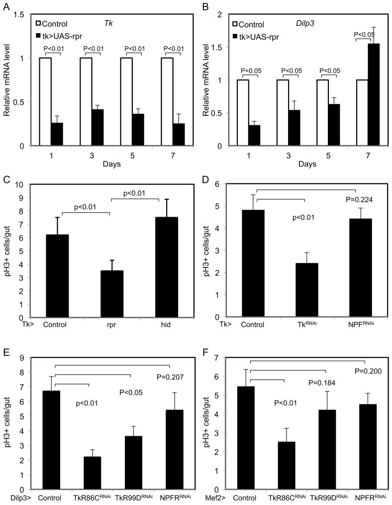
Intestinal stem cells in the adult Drosophila midgut are regulated by growth factors produced from the surrounding niche cells including enterocytes and visceral muscle. The role of the other major cell type, the secretory enteroendocrine cells, in regulating intestinal stem cells remains unclear. We show here that newly eclosed scute loss-of-function mutant flies are completely devoid of enteroendocrine cells. These enteroendocrine cell-less flies have normal ingestion and fecundity but shorter lifespan. Moreover, in these newly eclosed mutant flies, the diet-stimulated midgut growth that depends on the insulin-like peptide 3 expression in the surrounding muscle is defective. The depletion of Tachykinin-producing enteroendocrine cells or knockdown of Tachykinin leads to a similar although less severe phenotype. These results establish that enteroendocrine cells serve as an important link between diet and visceral muscle expression of an insulin-like growth factor to stimulate intestinal stem cell proliferation and tissue growth.


Figures

Figure 1
Figure 1. EE-less fly guts after loss of sc function have growth defects
(A) The number of Pros+ nuclei was counted within 0.08 mm2 surface area of a microscopic image from a similar region of each posterior midgut. The scRNAi midguts were completely devoid of EEs. For this and all other figures, data are presented as mean +/− SEM (error bar). (B) EE quantification in the midguts of flies with the genotypes indicated. Control was w-, the deficiency for sc was Df(1)sc10-1 and for ato was Df(3R)p13. Young flies were 7 days old and aged flies were 21 days old. NS is non-significant with P>0.05, and all P values are from Student’s t test. (C, D) Light microscope images of control and esg>scRNAi fly midguts. The arrow and hair line point to the posterior midgut region where images were taken to measure the diameter. (E) Quantification of the diameter of the midguts after starving (1% sucrose) or normal feeding condition. (F) The cross-section area of each enterocyte based on confocal images of Armadillo staining that outlined the cell shape was measured and the average is plotted as shown. (G, H) Midguts from fertilized females (7–10 days old) were homogenized and used for enzymatic assay. Each genotype corresponded to 5–6 samples of 10 midguts each. The sample with the highest activity in each enzyme assay was set as 100% and all others were calculated as a fraction.
Figure 2
Figure 2. EE-less guts have ISC proliferation and Dilp3 expression defects
(A–B) Newly hatched flies (day 1) were collected and kept in normal food vials or plastic vials with filter paper soaked with 1% sucrose (starved). Each day after, midguts were dissected from flies of the indicated genotypes and stained for p-H3 to detect mitotic cells. Average number of p-H3+ cells is plotted as shown. The esg>GFP in panel A or sc/+ in panel B served as controls. The deficiency is Df(1)sc10-1. (C) Dilp3 mRNA expression assayed by qPCR. Newly hatched esg>GFP (control) and esg>GFP, scRNAi flies were kept in normal food vials for 1 to 5 days as indicated. At each indicated day, 10 flies from each sample were used for gut dissection, RNA isolation and qPCR. Each qPCR cycle number of Dilp3 was normalized with that of rp49 in a parallel reaction of the same RNA sample. The lowest Dilp3 expressing sample esg>scRNAi at day 1 was set as 1 (first black bar) and all other samples were calculated as relative level and plotted as shown. (D–E) Dilp3 promoter-Gal4 driven UAS-GFP expression (Dilp3>GFP) illuminates the smooth muscle surrounding the adult midgut epithelium. This expression of muscle Dilp3>GFP is not altered when the UAS-scRNAi construct is also driven by this Dilp3 promoter. (F–K) Confocal images of midgut at an outer focal plane showing the visceral muscle staining, an inner focal plane showing the epithelium staining and 3D reconstruction of multiple focal planes. The control flies contained the combination of esg-Gal4 and Dilp3-Gal4 together driving UAS-GFP expression. The bottom panels I–K were from a fly strain that also contained the scRNAi construct.
Figure 3
Figure 3. Increasing the number of EEs promotes ISC division partly via Dilp3
(A–D) Down-shift experiments were performed by placing the esgGal4-tubulinGal80ts (esgts>) flies first at 29°C for 3 days and then back to room temperature (approximately 23°C). On the indicated days, fly guts were dissected to use for sc mRNA and phyllopod mRNA PCR assays. Each sc and phyllopod PCR cycle number was normalized with that of rp49, and the normalized control sample at each time point was set as 1 and the normalized UAS-sc sample of the same time point was calculated as fold change over the control. Equal numbers of flies from the same vials were used separately for Pros and p-H3 staining and quantification. (E) 3 days old flies kept at room temperature with the genotype esgts>GFP (control) and esgts>GFP, UAS-sc were shifted to 29°C for 3 days and then shifted back to room temperature for additional 3 days. The normalized Dilp3 mRNA of control at each time point was set as 1 and that of the UAS-sc of the same time point was calculated as fold change. (F) The tubulin-Gal4 driver with the temperature sensitive repressor tubulinGal80ts (tubulin, ts>) were crossed together with the transgenic constructs UAS-GFP as control, UAS-sc to increase the EE formation, and UAS-Dilp3RNAi to deplete the Dilp3 RNA. Flies approximately 7 days old were transferred to 29°C for 4 days to allow the expression of the transgenes. The flies were then used for gut dissection and p-H3 staining.
Figure 4
Figure 4. Tk secreting EEs have a role in regulating Dilp3 and ISC proliferation
(A–B) Tk-Gal4 flies were crossed with UAS-rpr to induce killing of a sub-population of EE cells. Tk>GFP was the control. Flies at the indicated days at room temperature after hatch were used for gut dissection and PCR assay. Each PCR cycle number of Tk and Dilp3 was normalized with the cycle number of rp49 in a parallel reaction of the same RNA sample. The control sample at each time point was set as 1 and UAS-rpr samples were plotted as a fraction of the control. (C) The same flies at 3 days as above and together with Tk>hid were used to quantify the number p-H3+ mitotic ISCs. (D) The flies containing the Tk-Gal4 driver were crossed with UAS-RNAi strains for Tk and NPF. The control was UAS-GFP. 3 days old progeny flies were dissected for p-H3 staining and quantification. (E–F) The flies containing the Dilp3-Gal4 or Mef2-Gal4 expressing in visceral muscle were crossed with UAS-RNAi strains for the receptors TkR86C, TkR99D and NPFR. The control was UAS-GFP. 3 days old progeny flies were dissected for p-H3 staining and quantification.

References

    1. Amcheslavsky A, Ito N, Jiang J, Ip YT. Tuberous sclerosis complex and Myc coordinate the growth and division of Drosophila intestinal stem cells. J Cell Biol. 2011;193:695–710. - PMC - PubMed
    1. Amcheslavsky A, Jiang J, Ip YT. Tissue damage-induced intestinal stem cell division in Drosophila. Cell Stem Cell. 2009;4:49–61. - PMC - PubMed
    1. Bardin AJ, Perdigoto CN, Southall TD, Brand AH, Schweisguth F. Transcriptional control of stem cell maintenance in the Drosophila intestine. Development. 2010;137:705–714. - PMC - PubMed
    1. Biteau B, Jasper H. EGF signaling regulates the proliferation of intestinal stem cells in Drosophila. Development. 2011;138:1045–1055. - PMC - PubMed
    1. Biteau B, Jasper H. Slit/Robo signaling regulates cell fate decisions in the intestinal stem cell lineage of Drosophila. Cell Rep. 2014;7:1867–1875. - PMC - PubMed

Publication types

LinkOut - more resources