. 2014 Oct 9;9(1):118-128.
doi: 10.1016/j.celrep.2014.08.042. Epub 2014 Sep 25.

SDF-1 inhibition targets the bone marrow niche for cancer therapy

Affiliations

SDF-1 inhibition targets the bone marrow niche for cancer therapy

Aldo M Roccaro et al. Cell Rep. .

Abstract

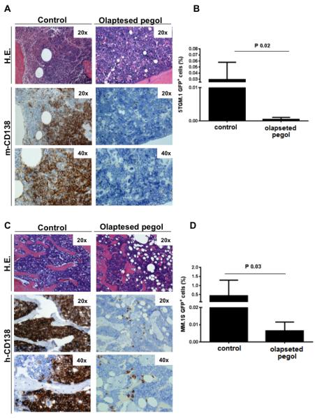
Bone marrow (BM) metastasis remains one of the main causes of death associated with solid tumors as well as multiple myeloma (MM). Targeting the BM niche to prevent or modulate metastasis has not been successful to date. Here, we show that stromal cell-derived factor-1 (SDF-1/CXCL12) is highly expressed in active MM, as well as in BM sites of tumor metastasis and report on the discovery of the high-affinity anti-SDF-1 PEGylated mirror-image l-oligonucleotide (olaptesed-pegol). In vivo confocal imaging showed that SDF-1 levels are increased within MM cell-colonized BM areas. Using in vivo murine and xenograft mouse models, we document that in vivo SDF-1 neutralization within BM niches leads to a microenvironment that is less receptive for MM cells and reduces MM cell homing and growth, thereby inhibiting MM disease progression. Targeting of SDF-1 represents a valid strategy for preventing or disrupting colonization of the BM by MM cells.


Figures

Figure 1
Figure 1. SDF-1 expression in metastasized bone marrow niches
(A) Bone marrow (BM) specimens were obtained from patients with multiple myeloma, MGUS, or solid tumors, and stained with anti-SDF-1 antibodies; MGUS samples were double-stained for SDF-1 (brown) and CD138 (blue). Normal BM was used as control. (B) SCID/Bg mice were injected (i.v.) with 5×10⁶ MM.1S-GFP+ cells; after 3 weeks, AlexaFluor(AF)633-conjugated anti-SDF-1α antibody (Ab) was administered i.v., and mouse skull BM niches were imaged after 4 hours, using in vivo confocal microscopy. Evans Blue was used to visualize blood vessels (MM.1S-GFP+/Luc+ cells: green; vessels: red; AF633-anti-SDF-1α Ab: blue). (C) Levels of SDF-1α in primary BM mesenchymal stromal cells isolated from MM patients (n=10) and healthy subjects (n=5) were evaluated by ELISA.
Figure 2
Figure 2. 193-G2-001/Olaptesed secondary structure and binding kinetics
(A) Pull-down binding assay of aptamer d-193-G2-001, using the biotinylated selection target d-SDF-1. Fitting with a 3 parameter algorithm revealed a Kd of 268 pM. (B) Secondary structure prediction for olaptesed, with potential base-pairs forming three hydrogen bonds depicted in red, and those forming two hydrogen bonds with each other shown in blue. (C) Biacore analysis of olaptesed pegol binding to immobilized human SDF-1α, at 37 °C under physiological buffer conditions. Raw data: black; fitted data: red.
Figure 3
Figure 3. In vitro characterization of ola-PEG
(A) CC, CXC, CX3C and XC chemokines (2 μM) were checked for competing with the binding of ola-PEG (12.5 nM) to immobilized human SDF-1α. As expected, mixtures of ola-PEG with SDF-1α or SDF-1β competed fully, but none of the other chemokines did so. Data are double-referenced, and are plotted as mean response units ± SD of n=2 injections. (B) Inhibition of SDF-1α-induced CXCR4 receptor internalization by ola-PEG. Jurkat cells were incubated with 0.3 nM SDF-1, plus various concentrations of ola-PEG. CXCR4 surface expression was quantified by flow cytometry, using a CXCR4 specific PE-labeled antibody. Data points are means ± SD for triplicate measurements. ola-PEG inhibits CXCR4 internalization with an IC50 of approximately 200 pM. (C) Inhibition of SDF-1-induced chemotaxis. Ola-PEG inhibits chemotaxis of Jurkat cells, with an IC50 of approximately 200 pM. Baseline level (fluorescence measured without SDF-1) is reached at approximately 800 pM ola-PEG. Means ± SD for triplicate measurements are shown. (D) Inhibition of SDF-1-induced CXCR7 activation by ola-PEG. The mean IC50 value obtained from three independent experiments was 5.1 nM.
Figure 4
Figure 4. Ola-PEG-dependent neutralization of SDF-1 reduces MM tumor progression in vivo
(A) Three weeks of pre-treatment with ola-PEG (20mg/kg injected every other day; s.c.) led to inhibition of MM tumor progression, shown with use of bioluminescence imaging. ola-treated mice are compared to mice that were pre-treated with AMD3100 (5,g/kg, daily; s.c.), or to untreated mice (n: 8/group). (B) Ola-PEG-pretreated mice presented with prolonged survival relative to AMD3100-pretreated or untreated mice (n: 10/group).
Figure 5
Figure 5. Ola-PEG-dependent neutralization of SDF-1 inhibits dissemination of MM cells from bone-to-bone
(A-D) ola-PEG-dependent inhibition of SDF-1 inhibited in vivo colonization of MM cells (GFP+5TGM1; GFP+MM.1S) - originating at a primary bone marrow site - to distant bone niches, as demonstrated by flow cytometry (GFP+) and immunostaining for murine (m) and human (h)-CD138. H.E. indicates hematoxylin-eosin staining (20×; 40×). Mice were euthanized when signs of paralysis were detected.
Figure 6
Figure 6. In vivo relevance of ola-PEG: effect on MM tumor growth
(A) SCID/Bg mice were injected (i.v.) with 5×10⁶ MM.1S-GFP+/Luc+ cells. Subsequently, mice were treated with vehicle (control), bortezomib (0.5 mg/kg, twice/week; i.p.) or ola-PEG (20 mg/kg, every other day; s.c.), alone or in combination (ola-PEG followed by Bortezomib) (n=5 per group). Tumor burden was detected by bioluminescence imaging, at different time points post-MM cell injection (t0: 2nd week; t1: 4th week; t2: 5th week). (B) Five weeks after MM cell inoculation, MM cell presence in BM structures of the skull was evaluated by intravital confocal microscopy (GFP+ MM cells: green; Evans Blue positive-blood vessels: red). Specific BM niches are highlighted, and relative magnification and 3D reconstruction are provided for each panel. (C) The presence of MM.1S-GFP+ cells ex vivo on femur tissues was detected by immunofluorescence. One representative image for one mouse from each group is shown (40×). For relative quantification, see Suppl. Figure 5E. (D) Primary MM BM-MSCs were treated with ola-PEG for 10 hours, and subsequently cultured in the presence of MM.1S cells for 8 hours. MM.1S cells were then harvested and cell lysates were subjected to Western blotting, with use of antibodies against p-ERK1/2, ERK1/2, p-cofilin, p-paxillin, p-Akt, p-Src, p-S6R, and p-GSK3. MM.1S cells cultured in the absence of MM BM-MSCs were used as control.

References

    1. Alsayed Y, Ngo H, Runnels J, Leleu X, Singha UK, Pitsillides CM, Spencer JA, Kimlinger T, Ghobrial JM, Jia X, et al. Mechanisms of regulation of CXCR4/SDF-1 (CXCL12)-dependent migration and homing in multiple myeloma. Blood. 2007;109:2708–2717. - PMC - PubMed
    1. Azab AK, Runnels JM, Pitsillides C, Moreau AS, Azab F, Leleu X, Jia X, Wright R, Ospina B, Carlson AL, et al. CXCR4 inhibitor AMD3100 disrupts the interaction of multiple myeloma cells with the bone marrow microenvironment and enhances their sensitivity to therapy. Blood. 2009;113:4341–4351. - PMC - PubMed
    1. Bleul CC, Fuhlbrigge RC, Casasnovas JM, Aiuti A, Springer TA. A highly efficacious lymphocyte chemoattractant, stromal cell-derived factor 1 (SDF-1) J Exp Med. 1996;184:1101–1109. - PMC - PubMed
    1. Bryden AA, Islam S, Freemont AJ, Shanks JH, George NJ, Clarke NW. Parathyroid hormone-related peptide: expression in prostate cancer bone metastases. Prostate Cancer Prostatic Dis. 2002;5:59–62. - PubMed
    1. Burwick N, Moreau AS, Jia X, Leleu X, Runnels J, Ngo HT, Melhem MR, Roccaro AM, Azab AK, Ghobrial IM. CXCR7 Regulates SDF-1 Induced Adhesion and Homing in Multiple Myeloma Blood. 2008;112 Abstract 1666. - PMC - PubMed

MeSH terms

Associated data