Maximum preservation of the media in carotid endarterectomy
- PMID: 25263623
- PMCID: PMC4533389
- DOI: 10.2176/nmc.tn.2014-0202
Maximum preservation of the media in carotid endarterectomy
Abstract
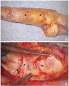
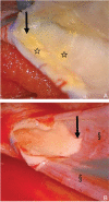
Carotid endarterectomy (CEA) is intended to remove atheromatous plaque by dissecting a plane between the intima and the media (circular medial fibers), but this may not be the optimal dissection plane. The present technique is based on identifying the plane that divides the media from the plaque, so preserving the media on the adventitia as much as possible. This plane is more difficult to find and follow than the easy-to-dissect plane usually located between the media and the adventitia, because the plaque invades the media and so the dividing plane is located within the media. In this prospective observational study, CEA was performed in 22 patients to histologically examine the excised plaques and small samples of the whole arterial wall, and evaluate the clinical outcomes. Plaque had invaded the luminal part of the media in the whole arterial wall sample of 80% of cases. Thin medial layers covering > 80% of the surface of the plaque were found in 16 of 22 plaques (73%). Some atheromatous component was sometimes left in the preserved media, rather than completely removed with the media. No morbidity or mortality had occurred by discharge. Only 1 small ipsilateral infarction (4.5%) and no restenosis of greater than 50% were detected during the mean follow-up period of 7 years. Since the plaque usually invades the media, the optimum dissection plane may be located within the media, dividing it into two layers. The presence of some remnant atheromatous components in the preserved media was not associated with surgical complications or restenosis.
Conflict of interest statement
The authors report no conflict of interest concerning the materials or methods used in this study or the findings specified in this article.
Figures


References
-
- Sacco RL, Adams R, Albers G, Alberts MJ, Benavente O, Furie K, Goldstein LB, Gorelick P, Halperin J, Harbaugh R, Johnston SC, Katzan I, Kelly-Hayes M, Kenton EJ, Marks M, Schwamm LH, Tomsick T, American Heart Association; American Stroke Association Council on Stroke; Council on Cardiovascular Radiology and Intervention; American Academy of Neurology : Guidelines for prevention of stroke in patients with ischemic stroke or transient ischemic attack: a statement for healthcare professionals from the American Heart Association/American Stroke Association Council on Stroke: co-sponsored by the Council on Cardiovascular Radiology and Intervention: the American Academy of Neurology affirms the value of this guideline. Stroke 37: 577– 617, 2006. - PubMed
-
- Bonati LH, Ederle J, McCabe DJ, Dobson J, Featherstone RL, Gaines PA, Beard JD, Venables GS, Markus HS, Clifton A, Sandercock P, Brown MM, CAVATAS Investigators : Long-term risk of carotid restenosis in patients randomly assigned to endovascular treatment or endarterectomy in the Carotid and Vertebral Artery Transluminal Angioplasty Study (CAVATAS): long-term follow-up of a randomised trial. Lancet Neurol 8: 908– 917, 2009. - PMC - PubMed
-
- Brott TG, Hobson RW, Howard G, Roubin GS, Clark WM, Brooks W, Mackey A, Hill MD, Leimgruber PP, Sheffet AJ, Howard VJ, Moore WS, Voeks JH, Hopkins LN, Cutlip DE, Cohen DJ, Popma JJ, Ferguson RD, Cohen SN, Blackshear JL, Silver FL, Mohr JP, Lal BK, Meschia JF, CREST Investigators : Stenting versus endarterectomy for treatment of carotid-artery stenosis. N Engl J Med 363: 11– 23, 2010. - PMC - PubMed
-
- Yadav JS, Wholey MH, Kuntz RE, Fayad P, Katzen BT, Mishkel GJ, Bajwa TK, Whitlow P, Strickman NE, Jaff MR, Popma JJ, Snead DB, Cutlip DE, Firth BG, Ouriel K, Stenting and Angioplasty with Protection in Patients at High Risk for Endarterectomy Investigators : Protected carotid-artery stenting versus endarterectomy in high-risk patients. N Engl J Med 351: 1493– 1501, 2004. - PubMed
-
- Moore WS: Technique of carotid endarterectomy, in Moore WS. (ed): Surgery for Cerebrovascular Disease, 2nd edition Philadelphia, WB Saunders, 1996, pp 368– 377
MeSH terms
LinkOut - more resources
Full Text Sources
Other Literature Sources

