

Arthroscopic Particulated Juvenile Cartilage Allograft Transplantation for the Treatment of Osteochondral Lesions of the Talus
- PMID: 25264516
- PMCID: PMC4175163
- DOI: 10.1016/j.eats.2014.06.004
Arthroscopic Particulated Juvenile Cartilage Allograft Transplantation for the Treatment of Osteochondral Lesions of the Talus
Abstract
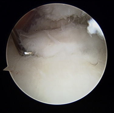
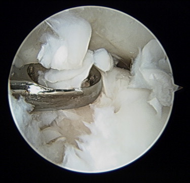
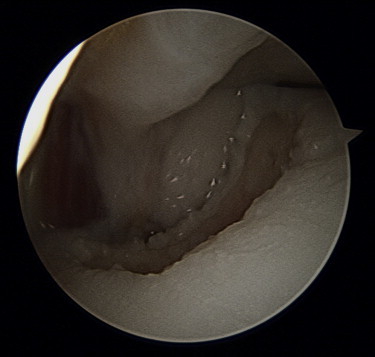
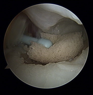
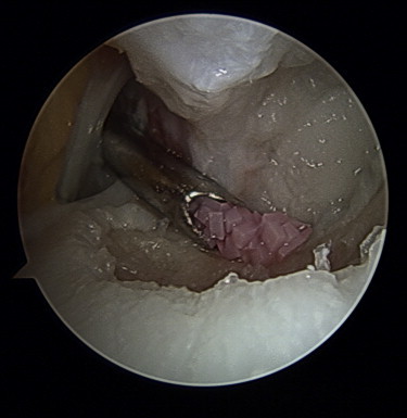
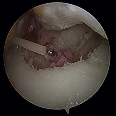
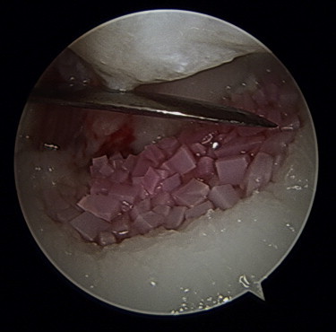
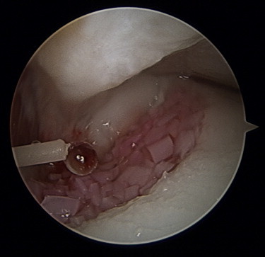
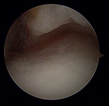
Several options exist for the treatment of osteochondral lesions of the talus. Particulated juvenile cartilage allograft transplantation (PJCAT) has become a viable treatment modality for osteochondral lesions of the talus that are not amenable to microfracture or for which microfracture has failed. Arthroscopic placement of this type of graft obviates the need for osteotomy or plafondplasty and does not prevent additional procedures from being performed through an anterior approach. Special instrumentation and setup are not required to perform this procedure. Our arthroscopic technique for placement of particulated juvenile cartilage into osteochondral lesions of the talus is described. Case series and outcomes after arthroscopic ankle PJCAT are currently not reported within the literature; however, it is believed that the outcomes are at least similar to those of open ankle PJCAT.
Figures
References
-
- Giurea A., DiMicco M.A., Akeson W.H., Sah R.L. Development-associated differences in integrative cartilage repair: Roles of biosynthesis and matrix. J Orthop Res. 2002;20:1274–1281. - PubMed
-
- Hunziker E.B., Kapfinger E., Müller M.E. Removal of proteoglycans from the surface of defects in articular cartilage transiently enhances coverage by repair cells. J Bone Joint Surg Br. 1998;80:144–150. - PubMed
-
- Qiu W., Murray M.M., Shortkroff S., Lee C.R., Martin S.D., Spector M. Outgrowth of chondrocytes from human articular cartilage explants and expression of α-smooth muscle actin. Wound Repair Regen. 2000;8:383–391. - PubMed
-
- Nehrer S., Spector M., Minas T. Histologic analysis of tissue after failed cartilage repair procedures. Clin Orthop Relat Res. 1999;365:149–162. - PubMed
-
- McGahan P.J., Pinney S.J. Current concept review: Osteochondral lesions of the talus. Foot Ankle Int. 2010;31:90–101. - PubMed
LinkOut - more resources
Full Text Sources
Other Literature Sources
