

Measurement of the glial fibrillary acidic protein and its breakdown products GFAP-BDP biomarker for the detection of traumatic brain injury compared to computed tomography and magnetic resonance imaging
- PMID: 25264814
- PMCID: PMC4394160
- DOI: 10.1089/neu.2014.3635
Measurement of the glial fibrillary acidic protein and its breakdown products GFAP-BDP biomarker for the detection of traumatic brain injury compared to computed tomography and magnetic resonance imaging
Abstract
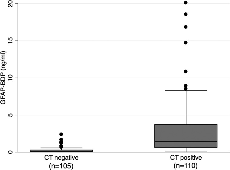
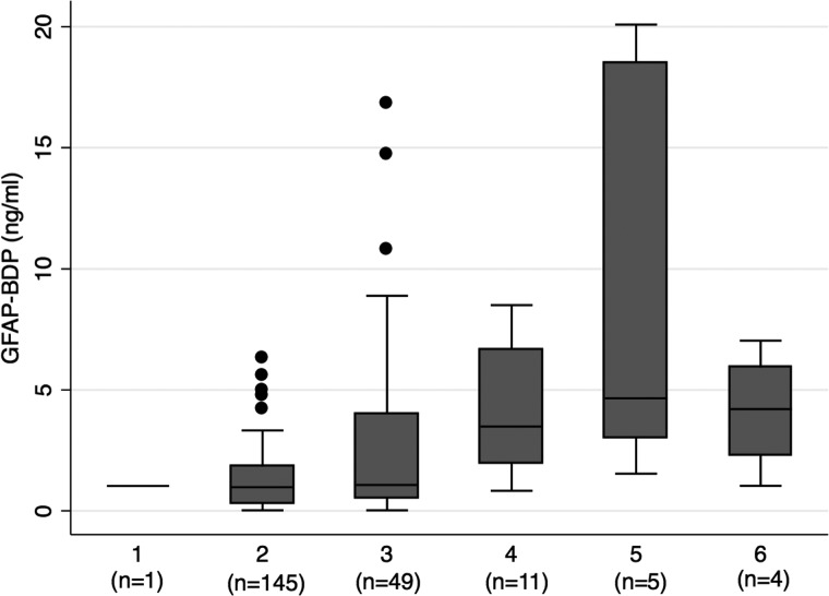
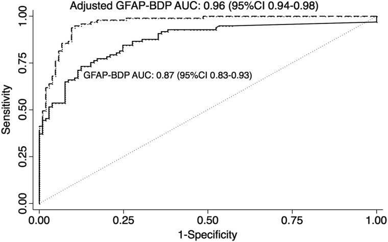
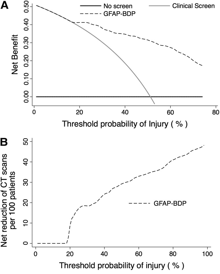
Glial fibrillary acidic protein and its breakdown products (GFAP-BDP) are brain-specific proteins released into serum as part of the pathophysiological response after traumatic brain injury (TBI). We performed a multi-center trial to validate and characterize the use of GFAP-BDP levels in the diagnosis of intracranial injury in a broad population of patients with a positive clinical screen for head injury. This multi-center, prospective, cohort study included patients 16-93 years of age presenting to three level 1 trauma centers with suspected TBI (loss of consciousness, post-trauma amnesia, and so on). Serum GFAP-BDP levels were drawn within 24 h and analyzed, in a blinded fashion, using sandwich enzyme-linked immunosorbent assay. The ability of GFAP-BDP to predict intracranial injury on admission computed tomography (CT) as well as delayed magnetic resonance imaging was analyzed by multiple regression and assessed by the area under the receiver operating characteristic curve (AUC). Utility of GFAP-BDP to predict injury and reduce unnecessary CT scans was assessed utilizing decision curve analysis. A total of 215 patients were included, of which 83% suffered mild TBI, 4% moderate, and 12% severe; mean age was 42.1±18 years. Evidence of intracranial injury was present in 51% of the sample (median Rotterdam Score, 2; interquartile range, 2). GFAP-BDP demonstrated very good predictive ability (AUC=0.87) and demonstrated significant discrimination of injury severity (odds ratio, 1.45; 95% confidence interval, 1.29-1.64). Use of GFAP-BDP yielded a net benefit above clinical screening alone and a net reduction in unnecessary scans by 12-30%. Used in conjunction with other clinical information, rapid measurement of GFAP-BDP is useful in establishing or excluding the diagnosis of radiographically apparent intracranial injury throughout the spectrum of TBI. As an adjunct to current screening practices, GFAP-BDP may help avoid unnecessary CT scans without sacrificing sensitivity (Registry: ClinicalTrials.gov Identifier: NCT01565551).
Keywords: biomarkers; imaging; traumatic brain injury.
Figures
References
-
- Stocchetti N., Pagan F., Calappi E., Canavesi K., Beretta L., Citerio G., Cormio M., Colombo A. (2004). Inaccurate early assessment of neurological severity in head injury. J. Neurotrauma 21, 1131–1140 - PubMed
-
- Vos P.E., Lamers K.J., Hendriks J.C., van Haaren M., Beems T., Zimmerman C., van Geel W., de Reus H., Biert J., and Verbeek M.M. (2004). Glial and neuronal proteins in serum predict outcome after severe traumatic brain injury. Neurology 62, 1303–1310 - PubMed
-
- Papa L., Lewis L.M., Silvestri S., Falk J.L., Giordano P., Brophy G.M., Demery J.A., Liu M.C., Mo J., Akinyi L., Mondello S., Schmid K., Robertson C.S., Tortella F.C., Hayes R.L., and Wang K.K. (2012). Serum levels of ubiquitin C-terminal hydrolase distinguish mild traumatic brain injury from trauma controls and are elevated in mild and moderate traumatic brain injury patients with intracranial lesions and neurosurgical intervention. J. Trauma Acute Care Surg. 72, 1335–1344 - PMC - PubMed
-
- Eng L.F., Ghirnikar R.S., and Lee Y.L. (2000). Glial fibrillary acidic protein: GFAP-thirty-one years (1969–2000). Neurochem. Res. 25, 1439–1451 - PubMed
-
- Lee Y.B., Du S., Rhim H., Lee E.B., Markelonis G.J., and Oh T.H. (2000). Rapid increase in immunoreactivity to GFAP in astrocytes in vitro induced by acidic pH is mediated by calcium influx and calpain I. Brain Res. 864, 220–229 - PubMed
Publication types
MeSH terms
Substances
Associated data
Grants and funding
LinkOut - more resources
Full Text Sources
Other Literature Sources
Medical
Research Materials
Miscellaneous
