. 2014 Sep 29;9(9):e107596.
doi: 10.1371/journal.pone.0107596. eCollection 2014.

Withaferin a alone and in combination with cisplatin suppresses growth and metastasis of ovarian cancer by targeting putative cancer stem cells

Affiliations

Withaferin a alone and in combination with cisplatin suppresses growth and metastasis of ovarian cancer by targeting putative cancer stem cells

Sham S Kakar et al. PLoS One. .

Abstract

Currently, the treatment for ovarian cancer entails cytoreductive surgery followed by chemotherapy, mainly, carboplatin combined with paclitaxel. Although this regimen is initially effective in a high percentage of cases, unfortunately within few months of initial treatment, tumor relapse occurs because of platinum-resistance. This is attributed to chemo-resistance of cancer stem cells (CSCs). Herein we show for the first time that withaferin A (WFA), a bioactive compound isolated from the plant Withania somnifera, when used alone or in combination with cisplatin (CIS) targets putative CSCs. Treatment of nude mice bearing orthotopic ovarian tumors generated by injecting human ovarian epithelial cancer cell line (A2780) with WFA and cisplatin (WFA) alone or in combination resulted in a 70 to 80% reduction in tumor growth and complete inhibition of metastasis to other organs compared to untreated controls. Histochemical and Western blot analysis of the tumors revealed that inclusion of WFA (2 mg/kg) resulted in a highly significant elimination of cells expressing CSC markers - CD44, CD24, CD34, CD117 and Oct4 and downregulation of Notch1, Hes1 and Hey1 genes. In contrast treatment of mice with CIS alone (6 mg/kg) had opposite effect on those cells. Increase in cells expressing CSC markers and Notch1 signaling pathway in tumors exposed to CIS may explain recurrence of cancer in patients treated with carboplatin and paclitaxel. Since, WFA alone or in combination with CIS eliminates putative CSCs, we conclude that WFA in combination with CIS may present more efficacious therapy for ovarian cancer.


Conflict of interest statement

Competing Interests: Co-author Surinder Batra is a PLOS ONE Editorial Board member. This does not alter the authors' adherence to all the PLOS ONE policies on sharing data and materials.

Figures

Figure 1
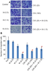
Figure 1. Effect of WFA and CIS both alone and in combination on cell migration.
A2780 cells were treated with different concentration of WFA and CIS both alone and in combination for 48 h. The cells were trypsinized and subjected for cell migration using Boyden chamber. Cells were stained with crystal violet and photographed (A). The stained cells were counted under microscope using three different areas; values shown are mean ± SD of three independent experiments. * Represents significant compared to control at p≤0.05 (B). Con  =  control, W  =  WFA. Values shown in parenthesis are µM.
Figure 2
Figure 2. Effect of WFA and CIS treatment on tumor growth.
A: 1×10⁶ A2780 cells were injected into female mouse ovary. After 10 days of post-injection, mice were treated with WFA and CIS both alone or in combination for four weeks. Mice were sacrificed; tumors were excised out, photographed and weighted. Tumors shown are representative from each group. B: Tumors weight was plotted from each group. Horizontal line represents median weight of each group. Treated group showed significantly lower weight than untreated mice. Results are mean (red line) and ± SD (vertical bar). * Represents significant compared to control at p≤0.05.
Figure 3
Figure 3. Effect of WFA and CIS both alone and in combination on tumor metastasis.
Mice were treated with WFA and CIS as indicated in Figure 2. Tumors and other tissues sections were stained with H&E and examined by a trained pathologist. Metastasis (shown by arrows) was observed in un-injected ovaries and livers and represent approximate 10% of the cells.
Figure 4
Figure 4. Immunohistochemical analysis of CD44 and CD34 positive cells in tumors collected from mock treated mice (control) and mice treated with WFA and CIS both alone and in combination.
The data shown is representative of two independent experiments. W  =  WFA. Values shown in parenthesis are mg/kg.
Figure 5
Figure 5. Immunohistochemical analysis of CD24 and CD117 positive cells in tumors collected from mock treated mice (control) and mice treated with WFA and CIS both alone and in combination.
The data shown is representative of two independent experiments. W  =  WFA. Values shown in parenthesis are mg/kg.
Figure 6
Figure 6. Immunohistochemical analysis of Oct4 positive cells in tumors collected from mock treated mice (control) and mice treated with WFA and CIS both alone and in combination.
The data shown is representative of two independent experiments. W  =  WFA. Values shown in parenthesis are mg/kg.
Figure 7
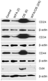
Figure 7. Western lot analysis of CD24, CD34, CD44, and Oct4 proteins from tumors collected from mock treated mice and mice treated with WFA and CIS both alone and in combination.
Beta-actin was used as an internal control. The data shown is representative of two independent experiments. Con  =  control, W  =  WFA. Values shown in parenthesis are mg/kg.
Figure 8
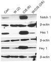
Figure 8. Western blot analysis of Notch1, Hes1 and Hey 1 proteins from tumors collected from mock treated mice and mice treated with WFA and CIS both alone and in combination.
Beta-actin was used as an internal control. The data shown is representative of two independent experiments. Con  =  control, W  =  WFA. Values shown in parenthesis are mg/kg.

References

    1. Siegel R, Naishadham D, Jemal A (2013) Cancer statistics. CA Cancer J Clin 63: 11–30. - PubMed
    1. Hunn J, Rodriguez GC (2012) Ovarian cancer: etiology, risk factors, and epidemiology. Clin Obstet Gynecol 55: 3–24. - PubMed
    1. National Comprehensive Cancer Network. NCCN Clinical Practice Guidelines in Oncology. Ovarian Cancer. Version 1.2013. Available: HTTP://www.nccn.org/professionals/physician_gls/pdf/ovarian.pdf
    1. Piccart MJ, Bertelsen K, Stuart G, Cassidy J, Mangioni C, et al. (2003) Long- term follow-up confirms a survival advantage of the paclitaxel-cisplatin regimen over the cyclophosphamide-cisplatin combination in advanced ovarian cancer. Int J Gynecol Cancer 13 Suppl 2: 144–148. - PubMed
    1. Matsuo K, Lin YG, Roman LD, Sood AK (2010) Overcoming platinum resistance in ovarian carcinoma. Expert Opin Investig Drugs 19: 1339–1354. - PMC - PubMed

Publication types

MeSH terms