. 2014 Sep 29;9(9):e107671.
doi: 10.1371/journal.pone.0107671. eCollection 2014.

Prohibitin-mediated lifespan and mitochondrial stress implicate SGK-1, insulin/IGF and mTORC2 in C. elegans

Affiliations

Prohibitin-mediated lifespan and mitochondrial stress implicate SGK-1, insulin/IGF and mTORC2 in C. elegans

Roxani Gatsi et al. PLoS One. .

Abstract

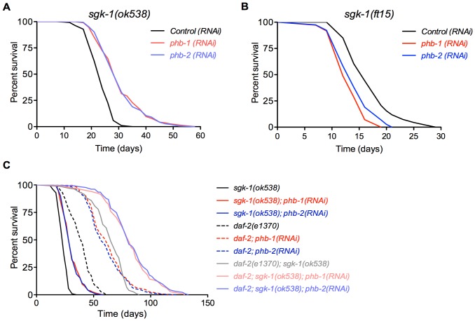
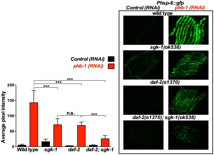
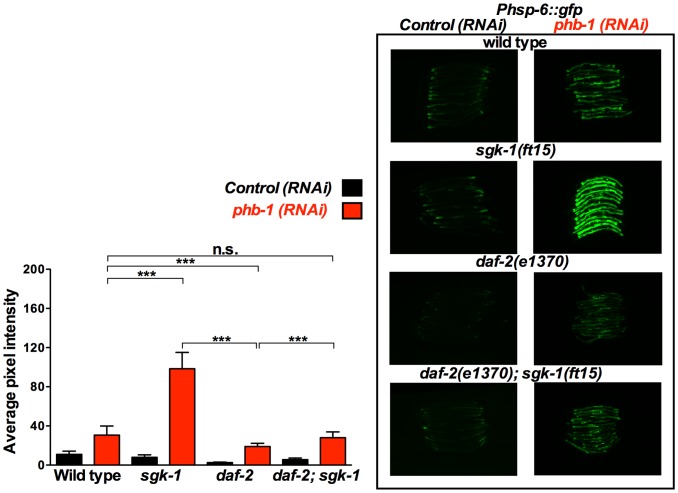
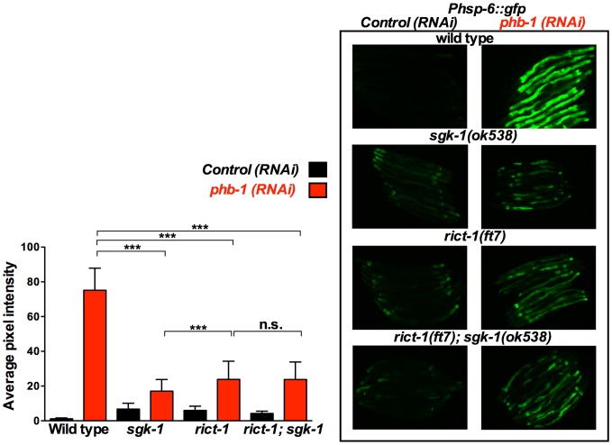
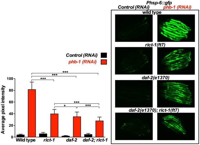
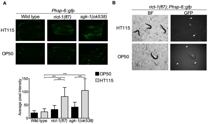
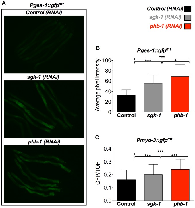
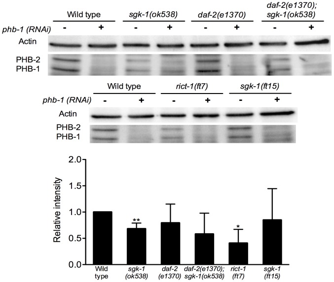
Lifespan regulation by mitochondrial proteins has been well described, however, the mechanism of this regulation is not fully understood. Amongst the mitochondrial proteins profoundly affecting ageing are prohibitins (PHB-1 and PHB-2). Paradoxically, in C. elegans prohibitin depletion shortens the lifespan of wild type animals while dramatically extending that of metabolically compromised animals, such as daf-2-insulin-receptor mutants. Here we show that amongst the three kinases known to act downstream of daf-2, only loss of function of sgk-1 recapitulates the ageing phenotype observed in daf-2 mutants upon prohibitin depletion. Interestingly, signalling through SGK-1 receives input from an additional pathway, parallel to DAF-2, for the prohibitin-mediated lifespan phenotype. We investigated the effect of prohibitin depletion on the mitochondrial unfolded protein response (UPRmt). Remarkably, the lifespan extension upon prohibitin elimination, of both daf-2 and sgk-1 mutants, is accompanied by suppression of the UPRmt induced by lack of prohibitin. On the contrary, gain of function of SGK-1 results in further shortening of lifespan and a further increase of the UPRmt in prohibitin depleted animals. Moreover, SGK-1 interacts with RICT-1 for the regulation of the UPRmt in a parallel pathway to DAF-2. Interestingly, prohibitin depletion in rict-1 loss of function mutant animals also causes lifespan extension. Finally, we reveal an unprecedented role for mTORC2-SGK-1 in the regulation of mitochodrial homeostasis. Together, these results give further insight into the mechanism of lifespan regulation by mitochondrial function and reveal a cross-talk of mitochondria with two key pathways, Insulin/IGF and mTORC2, for the regulation of ageing and stress response.


Conflict of interest statement

Competing Interests: The authors have declared that no competing interests exist.

Figures

Figure 1
Figure 1. SGK-1 interacts with prohibitins to regulate lifespan.
Lifespan curves are represented as the percentage of animals remaining alive against animal age (days). Combined lifespan data from independent experiments are shown in Table S1. A. Prohibitin depletion by RNAi against phb-1 or phb-2 at 20°C extends the lifespan of sgk-1(ok538) loss of function but not of akt-1(ok525), akt-2(ok393) or age-1(hx546) (see Figure S1 and Table S1). B. Knockdown of phb-1 or phb-2 shortens the lifespan of sgk-1(ft15) gain of function. C. Prohibitin knockdown extends the lifespan of daf-2(e1370) and of the daf-2(e1370); sgk-1(ok538), with the latest indicating additive effect.
Figure 2
Figure 2. sgk-1 and daf-2 mutants suppress the prohibitin depletion-mediated induction of the UPRmt reporter, Phsp-6::gfp.
Fluorescent microscopy of Phsp-6::gfp animals subjected to control RNAi (empty vector pL4440) or phb-1 RNAi (right panel) and graphical representation of the quantification of average pixel intensity under the corresponding conditions (left panel). Images were acquired under the same exposure, at the young adult stage. Prohibitin depletion at 20°C induced the UPRmt, as recorded by the mitochondrial chaperone reporter, Phsp-6::gfp. daf-2(e1370) and sgk-1(ok538) loss of function suppressed the prohibitin induced UPRmt. daf-2(e1370); sgk-1(ok538) double mutant caused an additive further suppression of the UPRmt, suggesting that daf-2 and sgk-1 are acting in parallel pathways to regulate the induction of the UPRmt upon prohibitin depletion. *** P value <0.0001, n.s. not statistically significant difference. Error bars denote SD. P values were calculated by using the student's t-test.
Figure 3
Figure 3. sgk-1 gain of function enhances the prohibitin depletion-mediated induction of the UPRmt in a daf-2 dependent manner.
Fluorescent microscopy of Phsp-6::gfp animals subjected to control RNAi (empty vector pL4440) or phb-1 RNAi (right panel) and graphical representation of the quantification of average pixel intensity under the corresponding conditions (left panel). Worms were imaged at the young adult stage under the same exposure. sgk-1(ft15) gain of function at 20°C enhanced the prohibitin induced UPRmt, as recorded by the mitochondrial chaperone reporter, Phsp-6::gfp. daf-2(e1370); sgk-1(ft15) suppressed the effect of the sgk-1 gain of function on the induction of the UPRmt upon prohibitin depletion, suggesting that daf-2 and sgk-1 are acting in parallel pathways to regulate the induction of the UPRmt upon prohibitin depletion. *** P value <0.0001, n.s. not statistically significant difference. Error bars denote SD. P values were calculated by using the student's t-test.
Figure 4
Figure 4. SGK-1 and RICT-1 act in the same pathway to regulate the prohibitin depletion-mediated induction of the UPRmt.
Fluorescent microscopy of Phsp-6::gfp animals subjected to control RNAi (empty vector pL4440) or phb-1 RNAi (right panel) and graphical representation of the quantification of average pixel intensity under the corresponding conditions (left panel). Worms were imaged at the young adult stage. sgk-1(ok538) and rict-1(ft7) loss of function at 20°C suppressed the prohibitin induced UPRmt as recorded by the mitochondrial chaperone reporter, Phsp-6::gfp. rict-1(ft7); sgk-1(ok538) did not cause further suppression of the UPRmt, suggesting that rict-1 and sgk-1 are acting in the same pathway to regulate the induction of the UPRmt upon prohibitin depletion. *** P value <0.0001, n.s. not statistically significant difference. sgk-1(ok538) and rict-1(ft7) mutants show increased Phsp-6::gfp expression when compared to wild type worms (sgk-1(ok538) vs. wild type: P<0.0001, rict-1(ft7) vs. wild type: P<0.0001, rict-1(ft7); sgk-1(ok538) versus wild type: P<0.0001). Error bars denote SD. P values were calculated by using the student's t-test.
Figure 5
Figure 5. rict-1 loss of function suppresses the prohibitin depletion-mediated induction of the UPRmt in a parallel pathway to daf-2.
Fluorescent microscopy of Phsp-6::gfp animals subjected to control RNAi (empty vector pL4440) or phb-1 RNAi (right panel) and graphical representation of the quantification of average pixel intensity under the corresponding conditions (left panel). Worms were imaged at the young adult stage. daf-2(e1370) and rict-1(ft7) loss of function at 20°C suppressed the prohibitin induced UPRmt as recorded by the mitochondrial chaperone reporter, Phsp-6::gfp. daf-2(e1370); rict-1(ft7) caused an additive further suppression of the UPRmt, suggesting that daf-2 and rict-1 are acting in parallel pathways to regulate the induction of the UPRmt upon prohibitin depletion. *** P value <0.0001, * P value <0.01, n.s. not statistically significant difference. Error bars denote SD. P values were calculated by using the student's t-test.
Figure 6
Figure 6. Induction of Phsp-6::gfp in sgk-1 and rict-1 mutants is food source dependent.
Fluorescent microscopy of wild type; Phsp-6::gfp, sgk-1(ok538); Phsp-6::gfp and rict-1(ft7); Phsp-6::gfp animals grown on either HT115 or OP50 bacteria. A. Fluorescent images of wild type; Phsp-6::gfp, sgk-1(ok538); Phsp-6::gfp and rict-1(ft7); Phsp-6::gfp young adults (upper panel) and graphical representation of the quantification of average pixel intensity under the corresponding conditions (lower panel). *** P value <0.0001, n = 25–35. Error bars denote SD. P values were calculated by using the student's t-test. B. Fluorescent stereoscope images of rict-1(ft7); Phsp-6::gfp animals growing on either HT115 or OP50 bacteria. Bright field (BF) and fluorescent images are shown. Arrowheads point to P0 animals and arrows to F1 animals. The induced expression the Phsp-6::gfp reporter is evident in the P0 generation and becomes very strong in the F1 generation of rict-1(ft7) animals grown on HT115 bacteria.
Figure 7
Figure 7. sgk-1 and phb-1 depletion by RNAi increases mitochondrial mass in the intestine and the body wall muscle.
Analysis of mitochondrial content in Pges-1::gfpmt and Pmyo-3::gfpmt animals treated with empty vector pL4440 (control RNAi), sgk-1 RNAi, or phb-1 RNAi. A. Fluorescent microscopy of Pges-1::gfpmt animals. Worms were imaged at day 1 of adulthood. B. Graphical representation of the quantification of average pixel intensity under the corresponding conditions. Prohibitin and sgk-1 depletion at 20°C increased intestinal mitochondrial mass as recorded by the intestinal mitochondrial reporter Pges-1::gfpmt. *** P value <0.0001, * P value <0.01. Error bars denote SD. P values were calculated by using the student's t-test. C. Graphical representation of the quantification of green fluorescence signal (GFP normalized to time of flight (TOF)) of Pmyo-3::gfpmt animals using the COPAS worm sorter. TOF and green fluorescence signal were recorded for each individual adult worm and was summarized by mean + SD. Worms were sorted at day 1 of adulthood. *** P value <0.0001, n = 100–200. Error bars denote SD. P values were calculated by using the student's t-test.
Figure 8
Figure 8. sgk-1 and rict-1 mutants have reduced levels of prohibitins.
Western blot analysis showing actin, PHB-1 and PHB-2 protein levels of wild type, sgk-1(ok538), daf-2(e1370), daf-2(e1370); sgk-1(ok538), rict-1(ft7) and sgk-1(ft15) animals treated with control RNAi () or phb-1 RNAi (+) (upper panel) and graphical representation of the quantification of PHB-1 average pixel intensity under the corresponding conditions normalized to the actin signal levels (lower panel). The data are represented as relative intensity normalized to the wild type control RNAi. PHB-1 and PHB-2 protein levels under phb-1 RNAi were not detectable for the quantification. The antibody raised against PHB-1 recognizes also PHB-2 , . The graph represents data from three independent experiments. Worms were grown at 20°C until young adult stage. sgk-1(ok538) and rict-1(ft7) mutants show a significant decrease in PHB-1 protein levels compared to the wild type control. daf-2(e1370), daf-2(e1370); sgk-1(ok538) and sgk-1(ft15) mutants do not have statistically significant different protein levels compared to wild type control. ** P value <0.05, *P value <0.1.

References

    1. Steglich G, Neupert W, Langer T (1999) Prohibitins regulate membrane protein degradation by the m-AAA protease in mitochondria. Mol Cell Biol 19: 3435–3442. - PMC - PubMed
    1. Nijtmans LG, de Jong L, Artal Sanz M, Coates PJ, Berden JA, et al. (2000) Prohibitins act as a membrane-bound chaperone for the stabilization of mitochondrial proteins. EMBO J 19: 2444–2451. - PMC - PubMed
    1. Coates PJ, Jamieson DJ, Smart K, Prescott AR, Hall PA (1997) The prohibitin family of mitochondrial proteins regulate replicative lifespan. Curr Biol 7: 607–610. - PubMed
    1. Back JW, Sanz MA, De Jong L, De Koning LJ, Nijtmans LG, et al. (2002) A structure for the yeast prohibitin complex: Structure prediction and evidence from chemical crosslinking and mass spectrometry. Protein Sci 11: 2471–2478. - PMC - PubMed
    1. Tatsuta T, Model K, Langer T (2005) Formation of membrane-bound ring complexes by prohibitins in mitochondria. Mol Biol Cell 16: 248–259. - PMC - PubMed

Publication types

MeSH terms